Download
1 / 49
490 likes | 648 Views
Peripheral nervous system. Nervous system. Central Nervous System brain spinal cord Peripheral Nervous System peripheral nerves cranial nerves spinal nerves. Division of PNS. Sensory Division picks up sensory information and delivers it to the CNS Motor Division

E N D
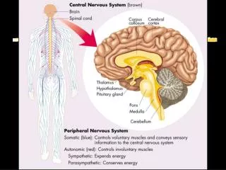
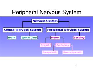
Nervous system • Central Nervous System • brain • spinal cord • Peripheral Nervous System • peripheral nerves • cranial nerves • spinal nerves
Division of PNS Sensory Division • picks up sensory information and delivers it to the CNS Motor Division • carries information to muscles and glands Divisions of the Motor Division • Somatic – carries information to skeletal muscle and skin • Autonomic – carries information to smooth muscle, cardiac muscle, and glands
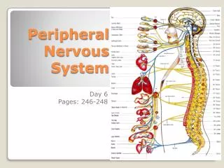
Spinal Cord • Surface structure • A. Meanings: • 1. Pia mater • 2. Arachnoid • 3. Dura mater • B. Spaces: • 1. Subarachnoid space - CSF fluid-filled, between arachnoid & pia mater • below end of spinal cord = lumbar cistern • 2. Epidural space - outside dura, between it and bone of spinal canal • - contains adipose tissue surrounding a venous plexus
Posterior Median Sulcus & Anterior Median Fissure Dorsal & Ventral median sulci midline grooves on their respective surfaces. a. anterior median fissure = largest of external grooves (a good orientation landmark) contains anterior spinal artery, within pia mater tissue b. Posterior / Dorsal median sulcus - more shallow Dorsal Septum = tissue extends from floor of sulcus deeper into cord Grooves
2. Posterolateral Septum & Anterolateral Fissure (dorsolateral & ventrolateral sulci) • where the dorsal and ventral roots respectively enter and exit the spinal cord. • 3. Posterior Intermediate Septum - only found in the cervical and thoracic cords • marks the separation of fibers carrying sensory information from the lower extremity more medial bundle = fasciculus gracilis • and fibers carrying sensory information from the upper extremity more lateral bundle = fasciculus cuneatus
Spinal Cord & Vertebral Segments • Spinal Cord has 31 pairs of spinal nerves exiting it: • 8 cervical • 12 thoracic • 5 lumbar 5 sacral • 1 coccygeal
Spinal column • Vertebral column has 26 bones: • 7 cervical (1=atlas; 2=axis) • 12 thoracic • 5 lumbar • 1 sacral (fused) • 1 coccyx (fused)
Spinal column & cord • Due to embryological growth pattern from ~ 3rd month on, spinal cord ends up shorter than the spinal canal, • a. cord segments don't match number of vertebral bodies at same level • b. cervical spinal nerves exit above the corresponding vertebral body (C8 above T1) • c. caudal to C8, spinal nerves exit below the corresponding vertebral body
Cord landmarks • 1.Conus Medullaris • By adulthood, as spinal column grows faster than cord • Spinal cord ends at level of lower border of L1 or at disk between L1 & L2 vertebral bodies • 2.Cauda Equina • (horse’s tail) - caudal to the conus medullaris • a. contains lumbosacral nerve roots - to the lower extremities • b. lumbar puncture or myelogram is given below level of L1 vertebra to avoid damage to the spinal cord. • 3. Filum Terminale • pia mater & neuroglia; - represents vestige of embryonic tail • a. tapered thin filament - from end of conus medullaris, runs through cauda equina • b. attached to dorsal surface of coccyx by coccygeal ligament (surrounding dural layer)
Cord landmarks • 4.Cervical & Lumbosacral Enlargements • accommodate the innervation of the upper and lower extremities • a. C4-T1: includes spinal nerves that make up brachial plexuses • b. L2-S3: lumbar & sacral plexuses • 5. Lumbar cistern • The subarachnoid space caudal to the end of the spinal cord, common site for lumbar puncture (lumbar tap)
Ventral & Dorsal Roots • 1. Dorsal roots break up into rootlets to enter cord all along dorsal surface at each level • 2. Ventral roots form by convergence of rootlets exiting from the ventral surface • 3. Dorsal Root Ganglion - found on the dorsal root just before it joins the ventral root • sensory nerve cell bodies (unipolar / pseudounipolar)
Spinal Nerve • formed by junction of dorsal & ventral roots, both sensory & motor • 1. Dorsal ramus: supplies innervation to skin, fascia, ligaments & deep muscles of back • 2. Ventral ramus: supplies ventral & lateral trunk as well as the limbs • also forms the plexuses to the extremities • 3.Rami Communicans: ventral branches of spinal nerve join to form sympathetic trunk • next to the vertebral body.
Dermatome • an area of skin that the sensory nerve fibers of a particular spinal nerve innervate
Sensory receptors • three groups • exteroceptive senses – senses associated with body surface; touch, pressure, temperature, pain • proprioceptive senses – senses associated with changes in muscles and tendons • visceroceptive senses – senses associated with changes in viscera
Autonomic Nervous System • functions without conscious effort • controls visceral activities • regulates smooth muscle, cardiac muscle, and glands • efferent fibers typically lead to ganglia outside CNS Two Divisions • sympathetic – prepares body for fight or flight situations • parasympathetic – prepares body for resting and digesting activities
Sympathetic Division • Origin from the intermediolateral cell column of thoracic and lumbar (L1 - L2 or L1 - L3) segments. • Some preganglionic fibers terminate in paravertebral ganglia.
Parasympathetic Division • Parasympathetic components derived from the brain and sacral segments of spinal cord.
Cranial source • Cranial nerve III (oculomotor nerve). Edinger-Westphal nucleus and ciliary ganglion • Facial nerve (VII): superior salivatory nucleus and submandibular ganglion • VII: Lacrimal nucleus and pterygopalatine ganglion • Glossopharyngeal nerve (IX): inferior salivatory nucleus and otic ganglion • Vagus nerve: Dorsal nucleus and ganglia in pulmonary plexus, plexuses in gastrointestinal tract • Sacral segment (S2 – S4) • Sacral autonomic nucleus
Neurotransmitters Cholinergic Fibers • release acetylcholine • preganglionic sympathetic fibers • preganglionic parasympathetic fibers • postganglionic parasympathetic fibers Adrenergic Fibers • release norepinephrine • postganglionic sympathetic fibers • Exceptions: • Sympathetic innervation of sweat gland is cholinergic
Receptors • Alpha • blood vessels of skin and internal organs, smooth muscle on pupil and internal sphincters • Constriction while alpha is stimulated • Beta • cardiac muscles, bronchus, blood vessels of skeletal muscles • Dilation while beta is stimulated • Muscarinic • end of all postganglionic parasympathetic nerve fibers • Slow, excitatory action • Nicotinic • between preganglionic and postganglionic neurons • Rapid, excitatory action
Blood Supply of the spinal cord • 1. two Posterior Arteries - together supply the dorsal horns and dorsal columns • 2. one Anterior Spinal Artery - supplies all other areas of the spinal cord • Both anterior and posterior spinal arteries branch off vertebral artery • 3. segmental spinal arteries (small) – branches from cervical vertebral artery, thoracic intercostals, and abdominal aorta