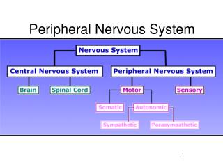
Peripheral Nervous System
Peripheral Nervous System. Chapter 13. Sensory Receptor Types. Nociceptors Respond to excess heat, pressure, or chemicals Tissue damage All parts of the body but brain Thermoreceptors Temperature of skin and blood Maintains homeostatic control via the hypothalmus Photoreceptors

Peripheral Nervous System
E N D
Presentation Transcript
Peripheral Nervous System Chapter 13
Sensory Receptor Types • Nociceptors • Respond to excess heat, pressure, or chemicals • Tissue damage • All parts of the body but brain • Thermoreceptors • Temperature of skin and blood • Maintains homeostatic control via the hypothalmus • Photoreceptors • Light absorbing pigments • Light detection
Sensory Receptor Types Mechanoreceptors Chemoreceptors • Touch, pressure, & vibrations • Bend or stretch PM of receptor cell = changing permeability • Stretch receptors – position of body parts • Hair cells -sound waves and H2O movements • Chemicals in the internal & external environment • O2 in arterioles • Osmoreceptors - changes in [blood solute] • Pheromone detection
Sensory Receptor Locations • Exteroceptors • Stimuli outside the body • Skin and special sense organs • Interoceptors • Stimuli within the body • Chemical messengers, tissue stretch, and temperature • Proprioceptors • Internal stimuli • Monitor position and stretch of joints, tendons, and muscles
Sensory Receptor Structures Unencapsulated Encapsulated • Free nerve endings • Most body tissues • Temperature and painful stimuli • Capsaicin and itch • Merkel discs • Deeper epidermal layers • Light touch • Hair follicle receptors • Shaft of hair follicle • Light touch and hair bending • Meissner’s corpuscles • Dermal papillae of sensitive and hairless skin • Discriminative touch • Pacinian corpuscles • Deep in the dermis • Deep pressure initially, vibration • Ruffini endings • Deep dermis and hypodermis • Deep continuous pressure • Muscle spindles • Perimysium of skeletal muscle • Detect muscle stretch & initiate a reflex • Golgi tendon • Insertion tendons • Activation inhibits contracting muscle
Sensory Input • All senses trigger the same TYPE of signal • Distinction occurs in activated brain area • Typically graded response, but AP’s possible • Sensory receptors detect sensations and carry to the brain • Awareness of environmental change • Brain constructs perceptions by integrating sensations with other information • Neuronal communication involving multiple brain areas
Sensory Adaptation • Sensory receptors become less responsive • Fewer action potentials • Limits reactions to normal background stimuli • Shower or hot tub temperature • Odors over time
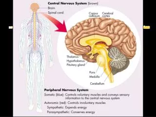
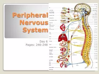
Decoding Nerves • PNS organ consisting of parallel bundles of axons enclosed by connective tissue layers • Epineurium • Perineurium • Endoneurium • Direction of transmission • Afferents: only to CNS • Dorsal root ganglia • Efferents: only from CNS • Sympathetic & parasympathetic ganglia • Mixed: carry both; most • Classified as cranial or spinal
Nerve Fiber Regeneration • Mature neurons don’t divide* • Cell body damage = death • Cut/compressed axons regenerate • Separated ends seal off and swell • Distal end of injury disintegrates • Lack of nutrients • Neurilemma maintained in endoneurium • Schwann cells proliferate & encourage axon growth • Guide ‘sprouting’ axons to original contacts • Greater distance decreases chances • Regrowth never exact = retraining • Extremely rare in CNS
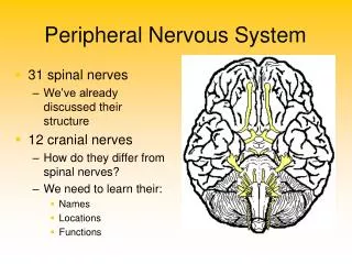
Cranial Nerves Ventral portion of the brain 1st 2 pairs attach to forebrain Remainders originate on brainstem
Testing Cranial Nerves for Disorders • Olfactory • Smell substances • Anosima • Optic • Eye chart • Anopsias • Oculomotor • Follow object; pupil reflex • Strabismus, double vision, ptosis • Trochlear • See oculomotor • Trigeminal • Close/move jaws; touch face with objects • Abducens • See oculomotor • Facial • Make various faces; tasting substances • Bell’s palsy, loss of taste, can’t close eye • Vestibulocochlear • Tuning fork; distance of sound • Deafness, vertigo, tinnitus • Glossopharyngeal • Swallowing & gag reflex; say ‘ah’ • Vagus • See glossopharyngeal • Horseness, swallowing problems, death • (Spinal) accessory • Move head/shoulders against resistance • Hypoglossal • Stick out, retract, & move tongue to sides
Spinal Nerve Anatomy • Roots: medial & afferent OR efferent • Dorsal root: peripheral receptors to spinal cord • Ventral root: ventral horn to skeletal muscles • Branches: laterally pass through intervertebral foramen • Dorsal ramus: dorsal trunk • Ventral ramus: limbs & rest of trunk • Meningeal branch: meninges and blood vessels • Plexus • Criss cross joining of ventral rami • Excludes T2 – T12
31 Pairs of Spinal Nerves • 8 cervical • Cervical plexus • Brachial plexus • 12 thoracic • Intercostal nerves & enlargements • 5 lumbar • Lumbar plexus • 5 sacral • Sacral plexus • 1 coccygeal • Tailbone & perineum
Cervical Nerves • Cervical plexus • Phrenic nerve: diaphragm • Irritation causes hiccups • Brachial plexus C5 – C8 • Median nerve: flexor muscles of the anterior forearm and small hand muscles • Carpal tunnel syndrome and suicide attempts • Radial nerve: extensor muscle of posterior forearm and triceps brachii • ‘Saturday night paralysis’ • Ulnar nerve: similar to median nerve • ‘Funny bone’ and paralysis/distortion of medial fingers
Lumbosacral Plexus • Innervates lower limbs, buttocks, and pelvic muscles • Lumbar plexus L1 – L4 • Femoral nerve: quadriceps and sartorius • Branches to saphenous • Obturator nerve: adductor muscles • Sacral plexus L4 – S4 • Sciatic nerve: entire lower leg (except anteriomedial thigh) • Tibial: hamstrings • Common fibular nerve: anterior tibialis
Reflex Arc • Receptor • Senses stimulus • Sensory neuron (afferents) • Message to the CNS • Integration center • Synapses in CNS • Monosynaptic (single motor or sensory neuron) • Polysynaptic (multiple interneurons) • Motor neuron (efferents) • Message to effectors • Effector • Muscle fibers or glands • Reflexes are rapid, predictable motor responses to a stimulus http://a248.e.akamai.net/7/248/430/20080327144023/www.mercksource.com/ppdocs/us/common/dorlands/dorland/images/arc_reflex%20a.(1).jpg
Classifying Reflexes • Somatic: activate skeletal muscle • Spinal: integration center is spinal cord • Stretch: ensures muscle length maintained (knee-jerk reflex) • Crossed extensor: withdrawl from painful stimuli (pin prick) • Superficial: cutaneous stimulation (plantar reflex) • Cranial nerve: integration center is brain stem • Corneal: stimulation causes blinking • Autonomic (visceral): activate smooth or cardiac muscle • Pupillary light: controls diameter of pupil (inside/outside) • Ciliospinal: ipsilateral pupil dilation from pain