Download
1 / 19
210 likes | 373 Views
The Serial Dilution Method of Bacteria Enumeration. Many studies require the quantitative determination of bacterial populations. The two most widely used methods for determining bacterial numbers are: The standard , or viable, plate count method and

E N D
Many studies require the quantitative determination of bacterial populations. The two most widely used methods for determining bacterial numbers are: • The standard, or viable, plate count method and • Spectrophotometer (turbid metric) analysis. • The standard plate count method is an indirect measurement of cell density ( live bacteria). • The spectrophotometer analysis is based on turbidity and indirectly measures all bacteria (cell biomass), dead and alive. Mohammed laqqan
The plate count (VIABLE COUNT) • The number of bacteria in a given sample is usually too great to be counted directly. • However, if the sample is serially diluted and then plated out on an agar surface in such a manner that single isolated bacteria form visible isolated colonies, the number of colonies can be used as a measure of the number of viable (living) cells in that known dilution. Mohammed laqqan
Keep in mind that if the organism normally forms multiple cell arrangements, such as chains, the colony-forming unit may consist of a chain of bacteria rather than a single bacterium. • In addition, some of the bacteria may be clumped together. Therefore, when doing the plate count technique, we generally say we are determining the number of Colony-Forming Units (CFUs) in that known dilution. • By extrapolation, this number can in turn be used to calculate the number of CFUs in the original sample. Mohammed laqqan
Normally, the bacterial sample is diluted by factors of 10 and plated on agar. After incubation, the number of colonies on a dilution plate showing between 30 and 300 colonies is determined. • A plate having 30-300 colonies is chosen because this range is considered statistically significant. Mohammed laqqan
If there are less than 30 colonies on the plate, small errors in dilution technique or the presence of a few contaminants will have a drastic effect on the final count. (too few to count (TFTC). • Likewise, if there are more than 300 colonies on the plate, there will be poor isolation and colonies will have grown together. (too numerous to count (TNTC). This plate has over 300 colonies and cannot be used for counting. This plate has between 30 and 300 colonies and is a suitable plate for counting. This plate less than 30 colonies and is unsuitable plate for counting. Mohammed laqqan
For a more accurate count it is advisable to plate each dilution in duplicate or triplicate and then find an average count. Fig 1 Mohammed laqqan Fig 2
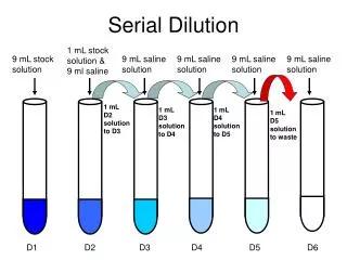
Procedure • 1. a- Take 6 dilution tubes, each containing 9.0 ml of sterile saline. Aseptically dilute 1.0 ml of a sample of E. colias shown inFig. 1 and described below. Mohammed laqqan
b. Insert the cotton-tipped end of the pipette into a blue 2 ml pipette filler. c. Flame the sample flask, insert the pipette to the bottom of the flask, and withdraw 1.0 ml (up to the "0" line; of the sample by turning the filler knob towards you. • Draw the sample up slowly so that it isn't accidentally drawn into the filler itself. • Reflame and cap the sample (see the Fig.). Using a Pipette to Remove Bacteria from a Tube Mohammed laqqan
Fig: 2 Mohammed laqqan
d. Flame the first dilution tube and dispense the 1.0 ml of sample into the tube by turning the filler knob away from you. Draw the liquid up and down in the pipette several times to rinse the pipette and help mix. Reflame and cap the tube. e. Mix the tube thoroughly by either holding the tube in one hand and vigorously tapping the bottom with the other hand or by using a vortex mixer (see Fig. 3). This is to assure an even distribution of the bacteria throughout the liquid and dissolve clumping of bacteria. Using a Vortex Mixer to Mix Bacteria Throughout a Tube Mohammed laqqan
Using the same procedure, aseptically withdraw 1.0 ml (see Fig. 2) from the first dilution tube and dispense into the second dilution tube. Continue doing this from tube to tube as shown in Fig. 1 until the dilution is completed. Mohammed laqqan
2. Using a new 1.0 ml pipette, aseptically transfer 0.1 ml (see Fig. 2)from each of the last three dilution tubes onto the surface of the corresponding plates of trypticase soy agar as shown in Figure 1 and Figure 4. Using a Pipette to Transfer Bacteria to an Agar Plate Mohammed laqqan
3. Using a turntable and sterile bent glass rod (see Fig. 5), immediately spread the solution over the surface of the plates as follows: a. Place the plate containing the 0.1 ml of dilution on a turntable. b. Sterilize the glass rod by dipping the bent portion in a dish of alcohol and igniting the alcohol with the flame from your burner. Let the flame burn out. Using a Bent Glass Rod and a Turntable to Spread a Bacterial Sample Mohammed laqqan
c. Place the bent portion of the glass rod on the agar surface and spin the turntable for about 30 seconds to distribute the 0.1 ml of dilution evenly over the entire agar surface. d. Replace the lid and resterilize the glass rod with alcohol and flaming. e. Repeat for each plate. Mohammed laqqan
Counting colonies • At the end of the incubation period, Count by looking at the bottom of the plate (while keeping the Petri plate closed). • Agar is translucent you should not have to open the plate • Select all of the Petri plates containing between 30 and 300 colonies. Count the colonies on each plate. • Plates with more than 300 colonies cannot be counted and are designated too many to count (TMTC). Also we might not have isolated colonies • Plates with fewer than 30 colonies are designated too few to count (TFTC). • If there are a lot of colonies on the plate helpful to use a marker to mark the colonies already counted Mohammed laqqan
CFU • Colonies forming units • Not the same as bacteria • 2 bacteria might have been very close and formed one colony • Calculate the number of bacteria (CFU) per milliliter or gram of sample by dividing the number of colonies by the dilution factor multiplied by the amount of specimen added to agar plate. • CFU per ml of sample = number of colonies / (amount plated X dilution) Mohammed laqqan
CFU calculation example • You count 46 colonies on your plate • You put 1 ml of bacterial culture into 99 ml of saline and plated 0.1 ml • Dilution 1/100 • CFU= 46 1/100 * 0.1 = 46 * 100 * 10 =46 000 Mohammed laqqan
End of lecture Mohammed laqqan