Download
1 / 75
800 likes | 1.16k Views
Osteogenesis imperfecta (OI) (Brittle bone/ Lobstein’s disease). Osteogenesis imperfecta is a heterogeneous group of heritable disorders characterized by impairment of collagen maturation which is manifested as congenital bone fragility

E N D
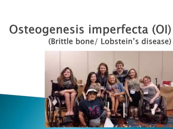
Osteogenesisimperfecta(OI)(Brittle bone/ Lobstein’s disease)
Osteogenesisimperfecta is a heterogeneous group of heritable disorders characterized by impairment of collagen maturation which is manifested as congenital bone fragility • Most common type of heritable bone disease (1/8000).
Type I collagen is less than normal or the quality is poorer due to defective intermolecular cross linkage of adjacent collagen molecules. • Type I collagen is an important component of bones, organ capsules, fascia, cornea, sclera, tendons, meninges, and dermis. • So the disease process results in widespread defects of these tissues
Causes • caused by mutations in the genes that codify for type I procollagen (ie, COL1A1 (chromosome 17) and COL1A2 (chromosome 7).). • inherited as dominant disorder or May show recessive pattern in some families from South Africa.
Types • 4 types of OI have been reported: • Type I - Mild forms • Type II - Severe • Type III – Moderate to Severe • Type IV -Mild to moderate
Extreme fragility & porosity of Bones leading to spontaneous fracture. However, these fractures heal readily but with a new bone of poor quality. • Due to repeated fractures person becomes deformed • Slight spinal curvature • Loose joints and Poor muscle tone • Early loss of hearing in some children • Tendency for bleeding
Blue sclera • Discolouration of the sclera usually giving them a blue-gray color. • The blue-gray color of the sclera is due to the underlying choroidal veins which show through. • This is due to the sclera being thinner than normal because of the defective Type I Collagen not forming correctly.
Blue sclera is also seen in • Osteopetrosis • Fetal rickets • Marfan syndrome • Ehlers-Danlos syndrome • Turner’s syndrome • Paget’s disease • Normal infants
Oral manifestations • Patient may present with dentinogenesisimperfecta- opalescent dentin • maxillary hypoplasia with class III malocclusion
Osteopetrosis • A group of rare hereditary skeletal defects of increased bone density resulting from defective remodeling due to failure in osteoclast function. • Endochondral, endosteal and periosteal growth without concomitant resorption results in thickening of bone.
Bone density progressively increases & marrow spaces are replaced by mineralized tissue. therefore patient develops anemia, thrombocytopenia & pancytopenia. • Dense bone is fragile with tendency to fracture. • Narrowing of foramens can cause compression of cranial nerves and related symptoms. • Radiographically bone appears sclerotic and dense.
Self limiting disease characterized by unusual cortical thickening of certain bones of infants • O/M- Mandible is commonly affected, residual defects can be seen even after the disease is cured, usually in the angle of mandible and ramus • R/F- Multifocal periosteal new bone formation- Onion peel appearance
Massive Osteolysis • Rare, idiopathic spontaneous progressive destruction of one or more bones and proliferation of vascular connective tissue resulting in total disappearance of the affected bone. • Radiographs shows radiolucency followed by fracture, dissolution and fragmentation of bone • H/P – Bone replaced by highly vascularized fibrous connective tissue
O/M • 30% maxillofacial involvement, usually the mandible. • Signs include mobile teeth, pain, malocclusion, midline deviation, pathologic fracture and obstructive sleep apnea. • Intramedullary lucency, loss of lamina dura, thinning of cortex. • Spontaneous arrest; radiation/surgery.
Achondrogenesis • Defective endochondral ossification • Life threatening condition
Achondroplasis • Disturbance in endochondral bone formation • Most common form of dwarfism • C/F- • Abnormally short stature with short limbs • Normal intelligence • Unusual strength so many adopt the occupation of professional wrestling
Chondro-ectodermal dysplasia(Ellis-Van Creveld syndrome) • Rare disease characterized by four components • Chondrodysplasia • Polydactyly • Ectodermal dysplasia affecting hair, teeth and nails • Congenital heart diseases • O/M- Fusion of middle portion of upper lip to maxillary gingival margin eliminating muco-buccal fold • Natal teeth, premature eruption of deciduous teeth, congenital absence of teeth particularly lower anteriors • Delayed tooth eruption, cone shaped teeth, enamel hypoplasia • Notching or submucosalclefting of alveolar process
Marfan syndrome • is a genetic disorder of the connective tissue. • Mutation of FBN1(Fibrillin 1) gene • The degree of manifestations vary in affected people. • Affected people have • Musculoskeletal, Cardiac ,ocular defects
Musculo-Skeletal abnormalities • Tall and slender built • Disproportionately long arms, legs and fingers - archnodactily- spidery fingers • A breastbone that protrudes outward or dips inward • A high, arched palate and crowded teeth • An abnormally curved spine(scoliosis) • Flat feet • flexible joints leading to hyper extensibility or dislocation.
Cardio-vascular manifestations • Aortic aneurysm. - Dilatation of aorta. • Aortic dissection – that weakens the vessel's structure and can result in a rupture, which may be fatal. • Valve malformations- malformed or overly elastic heart valves, eventually lead to heart failure. • Enlargement of proximal pulmonary artery
Ocular manifestations • Ectopialentis/ Lens dislocation. Due to weakening of supporting structures • Retinal detachment or tear • Early-onset glaucoma or cataracts.
Treatment • No cure for Marfan syndrome • treatment focuses on preventing the various complications
Did Abraham Lincoln have Marfan syndrome? • based on Lincoln's unusual physical appearance, Dr. Abraham Gordon proposed in 1962 that Lincoln had Marfan syndrome.
Craniosynostosis Syndromes • Group of disorders in which one or more of the sutures close too early, resulting in abnormal shape of head. • Premature closure of the sutures may also cause increase of intracranial pressure • Also the skull or facial bones change from a normal, symmetrical appearance.
Symptoms • a distorted skull shape • Open fontanel, or "soft spot" on the infant's skull • early disappearance of the fontanel • slower growth of head compared with the body • hard ridge forming along the suture, depending on the type of craniosynostosis
Simple- only one suture involved • Complex or compound- multiple sutures are involved • Syndromiccraniosynostosis
Craniofacial Dysostosis/ Crouzon syndrome • is primarily characterized by premature closure of the fibrous joints (cranial sutures) between certain bones in the skull (craniosynostosis) and distinctive facial abnormalities. • Cranial and facial malformations may vary, ranging from mild to potentially severe
Affected individuals often have • a prominent forehead (frontal bossing); • a curved nose due to deviated nasal septum • Narrowed or obliterated anterior nares- “parrot beak” like nose • unusually flat or underdeveloped mid-facial regions (mid-face hypoplasia) • a short upper lip. • hypoplastic maxilla with relative mandibularprognathism. • Clefting of the lip and/or palate
Dental findings • highly arched • narrow palate • crowded teeth • open bite/ • malocclusion.
Ocular changes • unusual shallowness of the orbit resulting in, proptosis (bulging of eyeballs) • exposure keratitis as well as conjunctivitis • Hypertelorism- eyes that are spaced apart wider than usual • Strabismus- Both eyes do not point in the same direction • loss in vision.
Apert’s syndrome • genetic disorder characterized by the premature fusion of certain skull bones (craniosynostosis) leading to abnormal shape of the head and face. • In addition, a varied number of fingers and toes are fused together (syndactyly).
Characteristic facial features • Midfacial hypoplasia-sunken appearance of midface • Proptosis and hypertelorism and vision problems • a beaked nose, • underdeveloped upper jaw leading to crowding of teeth and malocclusion.
Individuals with Apert syndrome have webbed or fused fingers and toes. • Polydactily - extra fingers or toes • hearing loss, • hyperhidrosis - unusually heavy sweating • oily skin with severe acne, patches of missing hair in the eyebrows • fusion of cervical vertebrae
Early fusion of the skull bones also affects the development of the brain, which can disrupt intellectual development. • Cognitive abilities in people with Apert syndrome range from normal to mild or moderate intellectual disability.


