Download
1 / 66
660 likes | 705 Views
Explore the intricate details of internal organs, including the digestive system, and the classification based on function, development, and constitutional types in the human body. Learn about the structure, functions, and topography of various visceral organs. Discover the vital components of the digestive system and their functions for metabolism.

E N D
The State Medical and Pharmaceutical University “Nicolae Testemiţanu” Viscera Functional Anatomy of the Digestive System Department of Human Anatomy Lecturer Dr. GlobaLilian
Plan • Viscera • Digestive System
Locomotor apparatus – movement • Internal organs - supply the locomotor apparatus
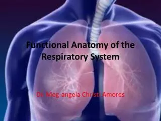
Classification of the internal organs According to functional point of view the viscera are divided into systems of organs and apparatuses. • Digestive system (energy, nutrients for growing up ) /the mouth, esophagus, gastrointestinal tract, liver and pancreas and salivary glands/; • Respiratory system (exchange of gases O2 to support burning)/(the nose, airways, larynx and lungs); • Urogenital system (excretion also skin)(the urinary and genital or reproductive organs) – (multiplication); • Controlling system Endocrine and Nervous systems (the ductless glands and cells which produce hormones); • Circulatory system (the heart and blood and lymph vessels); • Defense system (the blood, lymphatics and bone marrow);
The organs are divided into tubular (hollow) and parenchymatous organs. • Cavitaryorganshave a common tubular structure; • The wall of the cavitary organs consist of few layers: • The mucous coat ( tunica mucosa ) • The submucous layer ( telasubmucosa ) • The muscular coat (tunica muscularis ) • The serous coat (tunica serosa ), or the adventitious coat ( tunica adventitia )
The parenchymatousorgans consist of parenchyma and stroma. • The parenchyma, which is formed from specific elements, that assure the function of organs; • The stroma, which has a connective tissue origin sustains the parenchyma and leads the vessels and nerves.
Classification of the viscera dependent on their development, topography, structure, and functions • a) According to the development organs are divided into: • I. Organs derived from endoderm • II.Organs derived from somatic mesoderm • III. Organs derived from ectoderm
b) According to the topographical principle organs are divided into: • I. Organs of the head • II. Organs of the neck • III. Organs of the thoracic cavity • IV. Organs of the abdominal cavity • V. Organs of the pelvis.
Viscera and constitutional types of the human body • The size, shape and position of the organs and vessels depend on constitutional type. • In asthenics the viscera are smaller and have a lower position as they were ptotic. The lungs are longer, because the thoracic cage is longer. The heart has a vertical position and the aorta is narrow. The stomach has almost a vertical position as well as the loops of the small intestine. The liver, spleen and the pancreas and the kidney are small. • In hypersthenics the heart is relatively large and has almost a horizontal position and the aorta is large. The lungs are short. The stomach has a transverse position as well as the loops of the small intestine. The liver, spleen and the pancreas and the kidney are large. • The normosthenic type is an intermediate between the hypersthenic and asthenic type, and the organs have an intermediate position according to the characteristics accounted above.
Topography • Syntopy • Skeletotopy • Holotopy
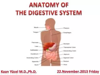
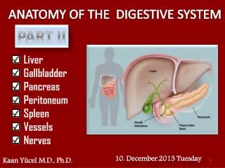
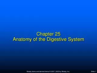
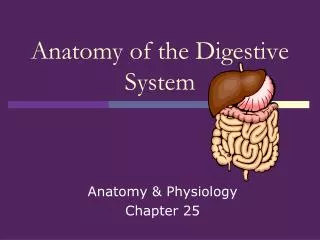
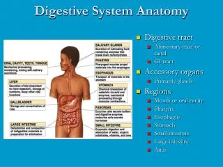
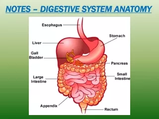
Digestive Sysytem the digestive system, or alimentary system (systema digestorium) is a complex of organs which provides mechanical and chemical treatment of food, absorption of the treated nutrients, and excretion of undigested remnants of the food.
Functions of the digestive system • Ingestion • Mechanical processing • Digestion • Secretion • Absorption • Excretion
Movement of digestive materials • Visceral smooth muscle shows rhythmic cycles of activity • Pacemaker cells • Peristalsis • Waves that move a bolus • Segmentation • Churn and fragment a bolus
Peristalsis Figure 24.4
Lips (labia) – protect the anterior opening • Cheeks – form the lateral walls • Hard palate – forms the anterior roof • Soft palate – forms the posterior roof • Uvula – fleshy projection of the soft palate
The tongue • primary functions include: • Mechanical processing • Assistance in chewing and swallowing • Sensory analysis by touch, temperature, and taste receptors
The pharynx • Common passageway for food, liquids, and air • Lined with stratified squamous epithelium • Pharyngeal muscles assist in swallowing • Pharyngeal constrictor muscles • Palatal muscles
Histology of the esophagus • Distinctive features of the esophageal wall include • Nonkeratinized, stratified squamous epithelium • Folded mucosa and submucosa • Mucous secretions by esophageal glands • A muscularis with both smooth and skeletal muscle portions • Lacks serosa • Anchored by an adventitia
Functions of the stomach • Bulk storage of undigested food • Mechanical breakdown of food • Disruption of chemical bonds via acids and enzymes • Production of intrinsic factor
Digestion and absorption in the stomach • Preliminary digestion of proteins • Pepsin • Permits digestion of carbohydrates • Very little absorption of nutrients • Some drugs, however, are absorbed • Mucous secretion containing several hormones • Enteroendocrine cells • G cells secrete gastrin • D cells secrete somatostatin
Histology of the stomach • Gastric glands • Parietal cells • Intrinsic factor, and HCl • Chief cells • Pepsinogen • Pyloric glands
Small intestine • Important digestive and absorptive functions • Secretions and buffers provided by pancreas, liver, gall bladder • Three subdivisions: • Duodenum • Jejunum • Ileum • Ileocecal sphincter • Transition between small and large intestine
Histology of the small intestine • Plicae • Transverse folds of the intestinal lining • Villi • Fingerlike projections of the mucosa • Lacteals • Terminal lymphatic in villus • Intestinal glands • Lined by enteroendocrine, goblet and stem cells
Small Intestine • Duodenal glands (Brunner’s glands) • produce mucus, buffers, urogastrone • Ileum • aggregated lymphoid nodules (Peyer’s patches)
Intestinal movements • Peristalsis • Segmentation • Gastroenteric reflexes • Initiated by stretch receptors in stomach • Gastroileal reflex • Triggers relaxation of ileocecal valve
The pancreas • Pancreatic duct penetrates duodenal wall • Endocrine functions • Insulin and glucagons • Exocrine functions • Majority of pancreatic secretions • Pancreatic juice secreted into small intestine • Carbohydrases • Lipases • Nucleases • Proteolytic enzymes
The Liver • Performs metabolic and hematological regulation and produces bile • Histological organization • Lobules containing single-cell thick plates of hepatocytes • Lobules unite to form common hepatic duct • Duct meets cystic duct to form common bile duct