Download

1 / 13
130 likes | 290 Views
Biological Background of Translation Process in E.Coli. Translation usually initiates at the AUG codon nearest to the 5´ end of the mRNA molecule. However, this does not happen in all cases.

E N D
Translation usually initiates at the AUG codon nearest to the 5´ end of the mRNA molecule. However, this does not happen in all cases. • There are some escape mechanisms that allow the initiation of translation at following, but still near the 5´ end, AUG codons: • leaky scanning: where the first AUG is bypassed due to inappropriate context. • Reinitiation, where translation initiates at an AUG codon before the correct initiation site and ends by reaching a stop codon. Translation reinitiates when the true AUG codon is found. • direct internal initiation: In this case the ribosome directly attaches near the true AUG codon without any scanning. • These mechanisms of the translation initiation process make more difficult the recognition of the TIS on a given genomic sequence.
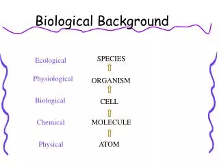
There are three different ways to read a given sequence in a given direction. Each of these ways of reading is referred to as reading frame. • The first reading frame starts at position 1, • The second starts at position 2, • The third starts at position 3. • The reading frame that is translated into a protein is named Open Reading Frame (ORF). A codon that is contained in the same reading frame with respect to another codon is referred to as “in-frame codon”. The coding region of an ORF is bounded by the initiation codon and the first in-frame stop codon. The coding region is surrounded by non-coding regions called 5´ and 3´ untranslated regions (UTRs).
The translation initiation region (TIR) in E.coli mRNA is characterized by the start codon and the Shine-Dalgarno base pairing of a region upstream of the gene's coding sequence with the 3'end of the 16SrRNA. • However, these domains are not sufficient to define an efficient TIR. Additional sequences and structures are required. How translation is affected by mRNA sequences upstream of the Shine- Dalgarno region or downstream of the start codon is not quite clear. • A nonrandom distribution of nucleotides in this region, revealed by statistical analysis, indicates that these sequences carry additional information in their primary structure for the efficiency of the initiation signals. Some scientists’ finding indicates that, nucleotides + 15 to +26, a sequence complementary to nucleotides 1471 to 1482 of the 16SrRNA, suggesting a second mRNA-rRNA base pairing contact besides the Shine-Dalgarno interaction.
If you are interested in this base pairing mechanism, please go to this website: http://www.pubmedcentral.nih.gov/picrender.fcgi? artid=330588&blobtype=pdf I think it could also become a target of your research. As for the initiation site prediction, I found some examples, such as this article named “Prediction of Translation Initiation Sites on the Genome of Synechocystis sp. Strain PCC6803 by Hidden Markov Model”. http://dnaresearch.oxfordjournals.org/cgi/content/abstract/4/3/179 Since I’m not that familiar with computer programming, this could only serve as a reference.
Translation Factors: • Initiation of translation begins with the 50s and 30s ribosomal subunits dissociated. • IF1 (initiation factor 1) blocks the A site to insure that the fMet-tRNA can bind only to the P site and that no other aminoacyl-tRNA can bind in the A site during initiation, • IF-2 is a small GTPase which binds fmet-tRNA and helps its binding with the small ribosomal subunit. • IF3 blocks the E site and prevents the two subunits from associating.
The 16s rRNA of the small 30S ribosomal subunit recognizes the ribosomal binding site on mRNA (the Shine-Dalgarno sequence, 5-10 base pairs upstream of the start codon(AUG)) The Shine-Delgarno sequence is found only in prokaryotes. This helps to correctly position the ribosome onto the mRNA so that the P site is directly on the AUG initiation codon. • IF-3 helps to position fmet-tRNA into the P site, such that fmet-tRNA interacts via base pairing with the mRNA initiation codon (AUG). Initiation ends as the large ribosomal subunit joins the complex causing the dissociation of initiation factors. • Prokaryotes (E. coli) can differentiate between a normal AUG (coding for methionine) and an AUG initiation codon (coding for formylmethionine and indicating the start of a new translation process).
Termination occurs when one of the three termination codons moves into the A site. These codons are not recognized by any tRNAs. Instead, they are recognized by proteins called release factors, namely • RF1 (recognizing the UAA and UAG stop codons) or • RF2 (recognizing the UAA and UGA stop codons). • RF-3 catalyzes the release of RF-1 and RF-2 at the end of the termination process. • These factors trigger the hydrolysis of the ester bond in peptidyl-tRNA and the release of the newly synthesized protein from the ribosome. • Example: If RF2 recognizes the stop codon UGA, translation is terminated after synthesis of a 25 amino acid peptide (which is degraded). If frameshifting occurs, the complete sequence of RF2 is synthesized.
The DNA sequence of the release factor 2 (RF2) gene revealed a stop codon at position 26 where the open reading frame switched to the +1 frame. Comparison of amino acid and mRNA sequences showed that ribosomes can shift reading frame at codon 25, avoid the stop codon, and decode the main portion of the message. Further work showed that this site-specific shift in reading frame is not just "noise" in translation but remarkably efficient: 30% of the ribosomes make complete RF-2 and 70% terminate at the zero frame UGA after 25 codons.
The RF-2 frameshift site is a "slippery" codon where the mRNA slides within the ribosome complex one nucleotide by breaking codon-anticodon pairing with peptidyl tRNA in the ribosome P site and re-establishing pairing with an overlapping codon in the new frame. In this case tRNA- Leu with anticodon 3'-GAG-5' pairs with CUU in the first frame and then with UUU in the +1 frame (CUU.UGA), inserting one leucine for four nucleotides in the mRNA.
Competition between termination and frameshifting is over which process captures the U of the UGA sequence. Release factor 2, the recoding product, promotes ribosome termination at UGA (and UAA) and so competes with recoding by enhancing termination at UGA, the 26th codon in the original frame. Competition is tilted in favor of recoding in two ways, The presence of a C 3' of the UGA makes a poor termination context, and CUU is a particularly shift-prone codon. If extra RF2 is present, frameshifting decreases so that less RF2 is made. When RF2 is in short supply termination loses, frameshifting wins, and RF2 concentration is restored.
The stimulatory signal is a short sequence three nucleotides 5' of the shift site that pairs with 16S rRNA of the translating ribosome, just like the Shine-Dalgarno pairing that occurs 5' of the AUG start codon during ribosome initiation. Thus, RF-2 mRNA has two Shine-Dalgarno sequences, one for initiation and one in the coding sequence for promoting frameshifting. Spacing between the Shine-Dalgarno sequence and the site of action is crucial and varies for these cases. With initiation the optimal spacing to the AUG is 5 bases, although there is considerable latitude (3 to 12 bases). With RF-2 required spacing is 3 bases. The implication is that pairing between mRNA and rRNA at these sites distorts the complex to either put the ribosome in an "initiation mode", a "plus mode" to force it forward, or a "minus mode" to force it backward.
BTW, I found an interesting article in this address: http://www.ncbi.nlm.nih.gov/books/bv.fcgi?rid=eurekah.section.19054 It indicated the mechnism of those translational factors, include initiation factors and release factors. For this article, it seems that these factors are related to the genome sequence of E. coli, for example, infA gene is responsible for IF1 synthesis. infA gene is located at 20 min on the E. coli chromosome. Therefore, in depth research would focus on these genes respectively, I wonder if you have time to finish these research before the deadline.