Μονάδα Εντατικής Θεραπείας Case reports
540 likes | 812 Views
Μονάδα Εντατικής Θεραπείας Case reports. Κάθε 3 η Τρίτη για όλο το ακαδημαικό έτος 2006-2007. Δομή παρουσίασης περιστατικού. Εισαγωγικό επιγραμματικό σχόλιο (δημογραφικά, αιτία / χρόνος / κατάσταση εισαγωγής στη ΜΕΘ) Παρούσα νόσος (από την αρχή) Ατομικό / οικογενειακό αναμνηστικό κλπ

Μονάδα Εντατικής Θεραπείας Case reports
E N D
Presentation Transcript
Μονάδα Εντατικής ΘεραπείαςCase reports Κάθε 3η Τρίτη για όλο το ακαδημαικό έτος 2006-2007
Δομή παρουσίασης περιστατικού • Εισαγωγικό επιγραμματικό σχόλιο(δημογραφικά, αιτία / χρόνος / κατάσταση εισαγωγής στη ΜΕΘ) • Παρούσα νόσος (από την αρχή) • Ατομικό / οικογενειακό αναμνηστικό κλπ • Εικόνα εισαγωγής στη ΜΕΘ – λίστα προβλημάτων (κατά συστήματα / εργαστηριακά ευρήματα) • Διαφορική διάγνωση / πρόγνωση • Επίκριση διαγνωστικών / θεραπευτικών ενεργειών με βάση τη βιβλιογραφία (Annotated references) • Έκβαση
Επίπεδα επιστημονικής εγκυρότητας ιατρικών γνώσεων • Level 1:Randomised, controlled trial with statistically significantresults • Level 2:Randomised, controlled trial with significantthreats to validity (e.g, small sample size, inappropriateblinding, weak methodology) • Level 3:Observational study with a concurrent controlgroup • Level 4:Observational study with a historical controlgroup • Level 5:Bench study, animal study, case series
Grade A Scientific evidence provided by randomised,well-designed, well-conducted, controlledtrials with statistically significant results thatconsistently support the guideline recommendation; Supportedby Level 1 or 2evidence
Grade Β Scientific evidence provided by well-designed, well-conducted observational studieswith statisticallysignificant results that consistentlysupport theguideline recommendation; Supported by Level 3 or 4 evidence
Grade C Scientific evidence from bench studies,animalstudies, case studies; Supported by Level5 evidence
Grade D Expert opinion provides the basis for the guidelinerecommendation, but scientific evidenceeither provided inconsistent results or waslacking
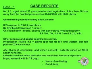
Εισαγωγικό σχόλιο Ασθενής, άντρας 77 ετών, 3η μέρα νοσηλείας στη ΜΕΘ, εισήχθη για υποστήριξη ζωτικών λειτουργιών με εικόνα καταπληξίας και αναπνευστικής ανεπάρκειας την 1η μετεγχειρητική μέρα μετά από εντερεκτομή για νέκρωση εντέρου στα πλαίσια αποφρακτικού ειλεού από συμφύσεις
Παρούσα νόσος • Αποφρακτικός ειλεός για 3 μέρες στο ΧΤ • Εικόνα οξείας κοιλίας • Χειρουργείο με ευρήματα ισχαιμίας – νέκρωσης λεπτού εντέρου • Εντερεκτομή και αναστόμωση • Μετεγχειρητικά, καταπληξία (ώρες) και αναπνευστική ανεπάρκεια • Μεταφορά στη ΜΕΘ / διασωλήνωση +12h
Προσωπικό / οικογενειακό ιστορικό • Περιτονίτιδα αδιευκρίνιστης αιτίας προ ετών • Αλκοολισμός? • Ισχαιμική καρδιοπάθεια με ΕΜ προ ετών?
Εικόνα εισαγωγής στη ΜΕΘ(προσέγγιση κατά συστήματα) 1. Όψη – θρέψη – δέρμα Όψη πάσχοντος Ψυχρά - υγρά άκρα Ακροκυάνωση Φυσιολογική θρέψη από την όψη
Εικόνα εισαγωγής στη ΜΕΘ(προσέγγιση κατά συστήματα) 2. ΚΝΣ / ΠΝΣ / ΑΝΣ Συγχυτικός – διεγερτικός Αντανακλάσεις νωθρές
Εικόνα εισαγωγής στη ΜΕΘ(προσέγγιση κατά συστήματα) 3. Καρδιαγγειακό SAP<80, HR> T <37 στην εισαγωγή CVP 5, Lac 8, ScvO2 22
Εικόνα εισαγωγής στη ΜΕΘ(προσέγγιση κατά συστήματα) 4. Αναπνευστικό pH 7.30, pO2 75, pCO2 29 (NRM) Extreme extraction
Εικόνα εισαγωγής στη ΜΕΘ(προσέγγιση κατά συστήματα) 5. Γαστρεντερικό Ειλεός
Εικόνα εισαγωγής στη ΜΕΘ(προσέγγιση κατά συστήματα) 6. Ουροποιητικό Ολιγουρία Κρεατινίνη 2.0mg/dl
Lab results (haematology) 27/10 12/11 21/11 WBC 6170 6100 1900 Neut % 60.9 82.1 77.5 Hgb 8.1 8.4 12.2 25.2 34.8 Hct 26.1 PLT 62000 153000 124000 Not done 1.10 INR 1.23
Lab results (biochemistry) 27/10 12/11 19/11 Glc 96 110 291 Urea 56 122 190 Creatinine 1.25 3.89 4.56 AST 36 42 45 LDH 1197 488 2036 11.2 Calcium Not done 11.55
Ερωτήματα προς συζήτηση • Ακολουθήθηκε η ορθή διαγνωστική προσπέλαση? • Έγινε έγκαιρη αντιμετώπιση / ανάταξη? • Ακολουθήθηκε η ενδεδειγμένη θεραπευτική? - Ανάταξη πίεσης - Ανάταξη άρδευσης - Ανάταξη ολιγουρίας - Ανάταξη αναπνευστικής ανεπάρκειας
“Christopher Reeve, our inspirational Chairman, passed away on October 10, 2004 from heart failure…” CNN News: “Reeve went into cardiac arrest Saturday at his home in Westchester County, New York, after developing aserious systemic infection during treatment for a pressure wound. He slipped into a coma and died Sunday afternoon at a hospital near his home…”
Septic Shock Sepsis: Defining a Disease Continuum Infection/Trauma SIRS Sepsis Severe Sepsis A clinical response arisingfrom a nonspecific insult, including 2 of the following: • Temp >38oC or < 36oC • HR > 90 beats/min • Respirations > 20/min • WBC >12,000/mm3or<4,000/mm3 or >10% immature neutrophils SIRS + presumed or confirmed infectious process Sepsis + Organ dysfunction • Cardiovascular (refractory hypotension) • Renal • Respiratory • Hepatic • Hematologic • CNS • Metab. acidosis SIRS = systemic inflammatory response syndrome. Bone et al. Chest. 1992;101:1644. Wheeler and Bernard. N Engl J Med. 1999;340:207.
A clinician, armed with the sepsis bundles, attacks the three heads of severe sepsis: hypotension, hypoperfusion and organ dysfunction. Crit Care Med 2004; 320(Suppl):S595-S597
6 hour Severe Sepsis/Septic Shock Bundle • Serum lactate measured. • Blood cultures obtained prior to antibiotic administration. • Broad-spectrum antibiotics administered within 3 hours of documented admission time. • In the event of hypotension (SBP < 90, MAP < 70) or lactate > 4 mmol/L, initial fluid resuscitation with 20-40 ml of crystalloid (or colloid equivalent) per estimated kg of body weight.
6 hour Severe Sepsis/Septic Shock Bundle • Vasopressors employed for hypotension during and after initial fluid resuscitation. • In the event of septic shock or lactate > 4 mmol/L, CVP and ScvO2 or SvO2 measured. • Inotropes (and/or PRBCs if hematocrit < 30%) delivered for ScvO2 <70 % or SvO2 < 65% if CVP > 8 mmHg.
24-Hour Severe Sepsisand Septic Shock Bundle • Glucose control maintained on average(or median) < 150 mg/dl (8.3 mmol/L) . • Drotrecogin alfa (activated) administered in accordance with hospital guidelines. • Steroids given for septic shock requiring continued use of vasopressors. • Adoption of a lung protective strategy with plateau pressures < 30 cmH2O for mechanically ventilated patients.
Patient enrollment and hemodynamic support Rivers et al. N Engl J Med 2001 SIRS and PAS < 90 mmHg or lactate > 4 Standard strategy n = 130 EGDT strategy n = 130 Randomisation n = 263 Vital and biological signs, ECG, SaO2, diuresis monitoring, arterial and venous catheterism CVP 8-12 mmHg CVP 8–12mmHg MAP > 65 mmHg Standard care ScvO2 monitoring and « agressive » strategy MAP > 65 mmHg Diuresis > 0.5 ml/kg/h Diuresis > 0.5 ml/kg/h ScvO2 > 70% 6 hours SaO2 > 93% Hte > 30% Clinical and biol. monitoring 8-72 hours Cardiac function Treatment < 6h n = 14 Treatment < 6h n = 13 VO2 Evaluation
Tissue perfusion is modified in sepsis • Vascular hyporeactivity and vasoplegia • inductible NOS • Catecholamine misuse • Endogenous (epinephrine, norepinephrine, vasopressine) • Exogenous • Perfusion heterogeneity • «SVR» is only a global estimation • Modifications in tissue distribution and/or use of O2 • Interstitial oedema (O2 diffusion abnormalities) • Hypoperfusion • Mitochondrial dysfonction
PiCCO™ System Central venous catheter Thermodilution femoral arterial catheter
Transpulmonary thermodilution Cardiac output measurement (vs Fick method) Tibby et al. Intensive Care Med 1997 9 CO Fick (L/min) n = 24 r2 = 0.99 y = 1.05x – 0.085 0 0 9 CO FATD (L/min)
SvO2 = SaO2 – [VO2 (Qc x Hb x 1.34)] ScVO2 monitoring in ICU Rivers et al. Current Opinion in Critical Care 2001; 7: 204-211 Factors that may influence SvO2 / ScvO2 value • Global indice of O2 extraction • if SaO2 = 1 SvO2 = 1 – EO2 • of no interest when severe hypoxemia and/or large variations of SaO2
Comparison of central-venous to mixed-venous oxygen saturation during changes in oxygen supply/demand Reinhart et al. Chest 1989; 95: 1216-1221 • It’s not the same value but : • Evolution is similar • Overestimation of SvO2 by ScvO2 • Same clinical signification • In ICU patients : • low ScvO2 < 60%= shock ou cardiac insifficiency • good correlation between ScvO2and SvO2 Reinhart et al. Int Care Med 2004
Vasopressors • Do not use low-dose dopamine for renal protection. Grade B Bellomo R, et al. Lancet 2000; 356:2139-2143
Vasopressors Vasopressin • Not a replacement for norepinephrine or dopamine as a first-line agent • Consider in refractory shock despite high-dose conventional vasopressors • If used, administer at 0.01-0.04 units/minute in adults • Grade E
Inotropic Therapy • Consider dobutamine in patients with measured low cardiac output despite fluid resuscitation. • Continue to titrate vasopressor to mean arterial pressure of 65 mm Hg or greater. Grade E
Inotropic Therapy • Do not increase cardiac index to achieve an arbitrarily predefined elevated level of oxygen delivery. Grade A Yu, et al. CCM 1993; 21:830-838 Hayes, et al. NEJM 1994; 330-1717-1722 Gattinoni, et al. NEJM 1995; 333:1025-1032
Sepsis-related Acute Renal Failure Mortality (%) p < 0.001 OSF at inclusion Neveu H. Nephrol Dial Transplant 1996
Dopamine “renal dose” • 58 published studies, n= 2149 • 18 RCT, n=864 • Mortality : 4,7 % vs 5,6 % RR 0,83 (0.39-1.77) • ARF : 15,3 % vs 19,5 % RR 0,79 (0.54-1.13) • RRT : 13,9 % vs 16,5 % RR 0,89 (0.66-1.21) Kellum J. Crit Care Med, 2001
Diuretics Mehta R. JAMA 2002 • 552 patients, 326 with diuretics • Prospective, obs. study • Impact of diuretics (OR, IC 95 %) Mortality : 1.65 (1.05-2.58) Dialysis dep.: 1.70 (1.14-2.53) • Uchino, Crit Care Med 2004 • Use of diuretics unrelated to excess risk of death in ICU • 54 centers, 23 countries, 1743 patients
Small differences in genotype make big differences to phenotype
Promoter Exon1 Intron1 Exon2 Quantity Change Quality Change Pathogen detection (CD14; MBL) Cytokines (TNF; IL-1;…) Coagulation (PAI-1; VII;…) Drug-metabolizing enzymes (CYP) … Pathogen detection (CCR5; TLR; MBL) FcgRII Coagulation Factors (Leyden;…) Drug-metabolizing enzymes … Relevant Polymorphisms
d2 Mice Susceptibility to Infection with Group A Streptococci 103 cfu Strepto Subcutaneous Goldman O. J Infect Dis 2003;187:854-61.
Community-Acquired Pneumonia and TNF polymorphisms 280 CAP No association with mortality rate LTa+250 AA genotype RR= 2.48 (1.28 – 4.78), Age-adjusted RR = 3.64 (1.28 – 10.66) Waterer GW. AJRCCM 2001; 163: 1599
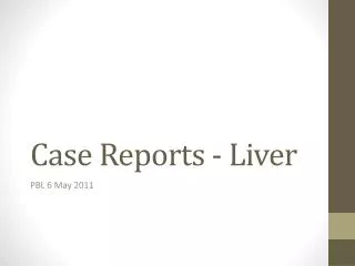
100 100 100 80 80 80 % Fatal Outcome 60 60 60 % Sepsis % MOF 40 40 40 20 20 20 13 19 29 4G/4G 4G/5G 5G/5G 4G/4G 4G/5G 5G/5G 4G/4G 4G/5G 5G/5G 4G/5G promoter polymorphism in the PAI-1 gene and severe trauma patients Menges, Lancet 2001;357:1096
Mechanical Ventilation ofSepsis-Induced ALI/ARDS • Reduce tidal volume over 1–2 hrs to 6 ml/kg predicted body weight • Maintain inspiratory plateau pressure < 30 cm H20 Grade B
Peak Airway Pressure Inspiratory Plateau Pressure PEEP (5 cm H2O 5 0