Bacteria Staining Procedures: Understanding Microbial Identification
670 likes | 714 Views
Explore the world of microbiology through staining procedures to identify bacteria based on their characteristics, atmospheric and nutritional requirements, colony morphology, and pathogenicity. Learn about Gram staining, acid-fast staining, and colony morphology analysis for microbial identification in the laboratory setting.

Bacteria Staining Procedures: Understanding Microbial Identification
E N D
Presentation Transcript
Domain BacteriaStaining Procedures • Three Categories of Staining Procedures • Simple stains • Structural staining procedures • Capsule stains • Spore stains • Flagella stains • Differential staining procedures • Gram and acid-fast staining procedures.
Domain BacteriaThe Gram Staining Procedure • Divides bacteria into 2 major groups: • Gram-positive (blue-to-purple) • Gram-negative (pink-to-red) • The final Gram reaction (positive or negative) depends upon the organism’s cell wall structure. • The cell walls of Gram-positive organisms have a thick layer of peptidoglycan, making it difficult to remove the crystal violet-iodine complex. • Gram-negative organisms have a thin layer of peptidoglycan, making it easier to remove the crystal violet; the cells are subsequently stained with safranin.
Various Gram-Positive Bacteria Streptococcus pneumoniae in blood culture. Chains of streptococci in smear from broth culture.
Gram-Negative Bacteria Loosely coiled Gram-negative spirochetes, Borrelia burgdorferi, the cause of Lyme disease. Gram-negative bacilli in a smear from a bacterial colony.
Domain BacteriaStaining Procedures (continued) • Some bacteria are neither consistently purple nor pink after Gram staining; they are known as Gram-variable bacteria; example Mycobacterium spp. • Mycobacterium spp. are often identified using the acid-fast stain. • The acid-fast stain • Carbol fuchsin is the red dye that is driven through the bacterial cell wall. • Heat is used to soften the waxes in the cell wall • Because mycobacteria are not decolorized by the acid-alcohol mixture, they are said to be acid-fast. • Ex TB
Domain BacteriaAcid-Fast Mycobacteria Many acid-fast mycobacteria in a liver biopsy. Acid-fast bacilli in a digested sputum specimen.
Domain BacteriaColony Morphology • A bacterial colony contains millions of organisms. • Colony morphology (appearance of the colony) varies from one species to another. • Colony morphology includes: size, color, overall shape, elevation and the appearance of the edge or margin of the colony. • Colony morphology also includes the results of enzymatic activity on various types of media. • As is true for cell morphology and staining characteristics, colony morphology is an important “clue” to the identification of bacteria.
Laboratory Identification of Pathogens • Obtain and label specimens from patients • Grow out bacterial cells • Isolate individual organisms • Multiply to form colonies • Stain cells so they can be seen (Gram Stain) • Perform tests to identify organisms
Methods of Identification Isolated colonies of bacteria growing on a solid medium
Size of colonies is determined by the organism’s generation time and is another important characteristic of a particular bacterial species. Formation of a bacterial colony on solid growth medium.
Factors Influencing Growth of Microorganisms • Light • Amount • Type • Temperature • Moisture • Food Availability • Atmosphere-Gas-Oxygen supply • pH
Domain BacteriaAtmospheric Requirements • Bacteria can be classified on the basis of their atmospheric requirements, including their relationship to O2 and CO2 • With respect to O2, bacterial isolates can be classified as: • Obligate aerobes- require 20-21% O2 • Microaerophilic aerobes -require O2 but less than 21% • Facultative anaerobes- can live and grow with or without molecular oxygen. Between 0-21% • Aerotolerant anaerobes- don’t require O2, grows better without O2, but can survive in areas with O2. • Obligate anaerobes- cannot survive in O2 environments • Capnophilic organisms grow best in the presence of increased concentrations of CO2 (5 to 10%).
Domain BacteriaNutritional Requirements • All bacteria need some form of the elements carbon, hydrogen, oxygen, sulfur, phosphorus and nitrogen for growth. • Some bacteria require special elements (e.g., calcium, iron or zinc). • Organisms with especially demanding nutritional requirements are said to be fastidious (“fussy”). • The nutritional needs of a particular organism are usually characteristic for that species and are sometimes important clues to its identity.
Domain BacteriaBiochemical and Metabolic Activities • As bacteria grow, they produce many waste products and secretions, some of which are enzymes. • Pathogenic strains of many bacteria, like staphylococci and streptococci, can be tentatively identified by the enzymes they secrete. • In particular environments, some bacteria produce gases such as carbon dioxide or hydrogen sulfide. • To identify bacteria in the lab, they are inoculated into various substrates (i.e., carbohydrates and amino acids) to determine whether they possess the enzymes necessary to break down those substrates.
Domain BacteriaPathogenicity • Many pathogens are able to cause disease because they possess capsules, pili, or endotoxins, or because they secrete exotoxins and exoenzymes that damage cells and tissues. • Frequently, pathogenicity is tested by injecting the organism into mice or cell cultures. • Examples of some common pathogenic bacteria: • Neisseria meningitidis, Salmonella typhi, Shigella spp., Vibrio cholerae, Yersina pestis, Treponema pallidum.
Bacterial By-Products • Endotoxins • Poison remains within the infected cell until it disintegrates • May cause typhoid fever & bacillary dysentery • Exotoxins • Poison is excreted by the cell into the surrounding area • May cause tetanus, gas gangrene, diphtheria and scarlet fever
Susceptibility • Now that the culture has been identified, we need to know what antibiotic will kill it. • The growth of the culture will have different antibiotics applied to determine which will kill the bacteria. • If the antibiotic does not kill the bacteria, the bacteria is called resistant. • Killed = susceptible to the antibiotic
Unique Bacteria • Rickettsias, chlamydias and mycoplasmas are bacteria, but do not possess all the attributes of typical bacterial cells. • Rickettsias and chlamydias have a Gram-negative type of cell wall and are obligate intracellular pathogens(i.e., they must live within a host cell). • Rickettsias do not grow on artificial culture media; they have “leaky membranes.” • Chlamydias are “energy parasites,” meaning they prefer to use ATP molecules produced by their host cell. Rickettsia prowazekii, the cause of epidemic louseborne typhus.
Unique Bacteria (continued) • Mycoplasmas • Smallest of the cellular microbes. • Lack a cell wall and therefore assume many shapes (they are pleomorphic). • In humans, pathogenic mycoplasmas cause primary atypical pneumonia and genitourinary infections. • Because they have no cell wall, they are resistant to drugs like penicillin that attack cell walls. • They produce tiny “fried egg” colonies on artificial media. SEM of Mycoplasma pneumoniae
Chlamydia • Energy parasites – use ATP from host • Gram negative cell wall • Obligate intracellular pathogen • Transmission is aerosol or direct contact • Can cause pneumonia, inclusion conjunctivitis, trachoma (blindness) as well as sexually transmitted infection.
Photosynthetic Bacteria • Photosynthetic bacteria include purple bacteria, green bacteria and cyanobacteria; they all use light as an energy source, but not in the same way. • Purple and green bacteria do not produce oxygen, whereas cyanobacteria do. • Photosynthesis that produces oxygen is called oxygenic photosynthesis. • Photosynthesis that does not produce oxygen is called anoxygenic photosynthesis.
The Domain Archaea • Archaea (meaning ancient) were discovered in 1977; they are procaryotic organisms. • Genetically, archaeans are more closely related to eucaryotes than they are to bacteria. • Archaeans vary widely in shape and live in extreme environments, such as extremely acidic, extremely hot and extremely salty environments. • Archaeans possess cell walls, but they do not have peptidoglycan (in contrast, all bacterial cell walls contain peptidoglycan).
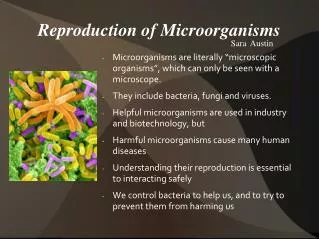
Categories of Microorganisms • Microorganisms can be divided into those that are truly cellular (bacteria, archaeans, algae, protozoa and fungi) and those that are acellular (viruses, viroids and prions). • Cellular microorganisms can be divided into those that are procaryotic (bacteria and archaeans) and those that are eucaryotic (algae, protozoa, and fungi). • Viruses, viroids and prions are often referred to as infectious agents or infectious particles.
Viruses Complete virus particles are called virions. Most viruses are from 10 to 300 nm in diameter. Viruses infect humans, animals, plants, fungi, protozoa, algae and bacterial cells. Some viruses, called oncogenic virusesor oncoviruses, cause specific types of cancer. A typical virion consists of a genome of either DNA or RNA, surrounded by a capsid (protein coat) which is composed of protein units called capsomeres. Some viruses (enveloped viruses) have an outer envelope composed of lipids and polysaccharides. Acellular Infectious Agents
Viruses have 5 properties that distinguish them from living cells: They possess either DNA or RNA – living cells possess both. They are unable to replicate on their own. Unlike cells, they do not divide by binary fission, mitosis or meiosis. They lack the genes and enzymes necessary for energy production. They depend on the ribosomes, enzymes and metabolites of the host cell for protein and nucleic acid production. Acellular Infectious Agents (continued)
Viruses are classified by: Type of genetic material (either DNA or RNA) Shape and size of capsid Number of capsomeres Presence or absence of an envelope Type of host it infects Disease it produces Target cell Immunologic/antigenic properties Acellular Infectious Agents (continued)
Herpesviruses acquiring their envelopes as they leave a host cell’s nucleus by budding.
Comparative sizes of virions, their nucleic acids and bacteria.
Viruses Origin of Viruses One theory states that viruses represent ancient derivatives of degenerate cells or cell fragments. Most scientists agree that viruses are nonliving entities. Bacteriophages Viruses that infect bacteria are known as bacteriophages or simply, phages. Virulent bacteriophagesalways cause what is known as the lytic cycle, which ends with the destruction of the bacterial cell. Temperate (lysogenic) bacteriophages do not cause the lytic cycle, but the DNA integrates into the bacterial chromosomes Acellular Infectious Agents (continued)
A partially lysed cell of Vibrio cholerae with attached virions of phage CP-T1.
The bacteriophage T4 is an assembly of protein components. Viral DNA enters the cell through the core. 20 facets, filled with DNA
The steps in multiplication of viruses are: Attachment Penetration Biosynthesis -when the viral nucleic acids take over the host cell and dictates what is produced Assembly -fitting the viral pieces together to produce complete virons Release Inclusion bodiesare remnants or collections of viruses; often seen in infected cells and used as a diagnostic tool to identify particular viral diseases. Acellular Infectious Agents (continued)
Infectious Nature of Viruses • Contact can be airborne or contact • Virus settles into/ onto the body and enters an open portal. • *Virus attaches to cell membrane, injects nucleic acids, “cons” cell’s DNA into synthesizing the new material. New viruses are made until the cell bursts (lyses), sending more viral material into the interstitial space. The process repeats
Acellular Infectious Agents (continued) • Latent Virus Infections • Viral infections in which the virus is able to hide from a host’s immune system by entering cells and remaining dormant. • Herpes viral infections are examples. • Once acquired, herpes virus infections (e.g., those that cause cold sores, genital herpes, and chickenpox/shingles) never completely go away; for example, chickenpox may be followed, years later, by shingles - both the result of the same virus.
Acellular Infectious Agents (continued) • Antiviral Agents • Antibiotics are not effective against viral infections. • Antiviral agents are drugs that are used to treat viral infections. • These agents interfere with virus-specific enzymes and virus production by disrupting critical phases in viral multiplication or inhibiting synthesis of viral DNA, RNA, or proteins.
Acellular Infectious Agents (continued) • Oncogenic Viruses or Oncoviruses • Viruses that cause cancer. • Examples include Epstein-Barr virus, human papillomaviruses and HTLV-1. • Human Immunodeficiency Virus (HIV) • The cause of acquired immunodeficiency syndrome (AIDS). • It is an enveloped, double-stranded RNA virus. • The primary targets for HIV are CD4+ cells.
Retroviruses • HTLV virus (RNA) – Leukemia • RNA tumor virus – Tumors • HIV (main one of this generation) – AIDS
Acellular Infectious Agents (continued) • Viroids and Prions (smaller and less complex infectious agents than viruses) • Viroids • Viroids are short, naked fragments of single-stranded RNA, which can interfere with the metabolism of plant cells. • Viroids are transmitted between plants in the same manner as viruses. • Examples of plant diseases caused by viroids: potato spindle tuber, and citrus exocortis.
Acellular Infectious Agents (continued) • Prions • Prions are small infectious proteins that cause fatal neurologic diseases in animals; examples: Scrapie, Bovine Spongiform Encephalopathy (“Mad Cow Disease”) and Creutzfeldt-Jacob disease. • Of all pathogens, prions are the most resistant to disinfectants. • The mechanism by which prions cause disease remains a mystery.
AlgaeCharacteristics and Classification • Algae are photosynthetic, eucaryotic organisms. • All algal cells consist of cytoplasm, a cell wall (usually), cell membrane, a nucleus, plastids, ribosomes, mitochondria and Golgi bodies. • Some have a pellicle, a stigma and/or flagella • Algae range in size from unicellular microscopic organisms (e.g., diatoms) to large, multi-cellular organisms (e.g., seaweeds or kelp). • Algae produce their energy by photosynthesis. • Some may use organic nutrients.
Algae: Medical Significance • One genus of algae, Prototheca, is a very rare cause of human infections. • Causes protothecosis. • Algae in several other genera secrete substances called phycotoxins. • Poisonous to humans, fish, and other animals. • If ingested by humans, the phycotoxins produced by the dinoflagellates can lead to paralytic shellfish poisoning.
Protozoa are nonphotosynthetic, eucaryotic organisms. Most protozoa are unicellular and free-living, found in soil and water. Most protozoa are more animal-like than plant-like. All protozoal cells possess a variety of eucaryotic structures/organelles. Protozoa cannot make their own food; they ingest whole algae, yeasts, bacteria and smaller protozoa for nutrients. ProtozoaCharacteristics
Protozoa do not have cell walls, but some possess a thickened cell membrane called a “pellicle,” which serves the same purpose – protection! A typical protozoan life cycle has 2 stages – a trophozoite and a cyst. The trophozoite is the motile, feeding, dividing stage. The cyst is the dormant, survival stage. Some protozoa are parasites. Parasitic protozoa cause many human diseases, such as malaria, giardiasis and trypanosomiasis. ProtozoaCharacteristics (continued)
Typical Pond Water Algae and Protozoa Voticella sp. Volvox sp. Stentor sp. Euglena sp. Paramecium sp. Amoeba sp.
ProtozoaCharacteristics (continued) • Protozoa are divided into groups, based on their method of locomotion: • Amebae move by means of pseudopodia (“false feet”) – example: Entamoeba histolytica, the cause of amebic dystentery. • Flagellates move by means of whiplike flagella – example: Giardia lamblia, the cause of giardiasis. • Ciliates move by means of hairlike cilia – example: Balantidium coli, the cause of balantidiasis. • Sporozoa have no visible means of locomotion – example: Plasmodium spp., which cause malaria.
Giardia lamblia – a Flagellate Drawing of Giardia lamblia trophozoite TEM of Giardia lamblia trophozoite
FungiCharacteristics • The study of fungi is called mycology. • Fungi are found virtually everywhere. • Some fungi are harmful, some are beneficial. • Fungi represent a diverse group of eucaryotic organisms that include yeasts, molds and mushrooms. • Fungi are the “garbage disposers” of nature. • Fungi are not plants – they are not photosynthetic!