Microbiology
Microbiology . The study of really small organisms . Microorganisms . Bacteria Fungi Archaea Protists Viruses and prions are not considered micro organisms – also studied . Who Studies Microbiology Microbiologist . Microbes can cause disease


Microbiology
E N D
Presentation Transcript
Microbiology The study of really small organisms
Microorganisms • Bacteria • Fungi • Archaea • Protists • Viruses and prions are not considered micro organisms – also studied
Who Studies MicrobiologyMicrobiologist • Microbes can cause disease • It is from the study of these diseases we can develop antibiotics to fight bacteria • Vaccines to fight viruses • Antifungal drugs to fight fungi • BUT: Microbes can be good • Beneficial in health • Nutrient recycling • Industrial production
THE Whose Who • Anton van Leuwenhock • Galileo • Louis Pasteur • Ferdinand Cohn • Robert Koch • MartinusBeijernick • Sergei Winogradsky
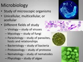
Areas of Microbiology • Microbial Physiology • Microbial Genetics • Medical Microbiology • Veterinary Microbiology • Environmental Microbiology • Evolutionary Microbiology • Industrial Microbiology • Aeromicrobiology • Food Microbiology • Pharmaceutical Microbiology
The Microscope http://www.bing.com/images/search?q=microscope+and+its+parts+and+functions&qpvt=microscope+and+its+parts+and+functions&FORM=IGRE&adlt=strict#view=detail&id=8AB8606B877ADDCAD6714845194BF254F1094E22&selectedIndex=7
Types of Microscopes • Since microorganisms are invisible to the unaided eye, the essential tool in microbiology is the microscope. One of the first to use a microscope to observe microorganisms was Robert Hooke, the English biologist who observed algae and fungi in the 1660s. In the 1670s, Anton van Leeuwenhoek, a Dutch merchant, constructed a number of simple microscopes and observed details of numerous forms of protozoa, fungi, and bacteria. During the 1700s, microscopes were used to further elaborate on the microbial world, and by the late 1800s, the sophisticated light microscopes had been developed. The electron microscope was developed in the 1940s, thus making the viruses and the smallest bacteria (for example, rickettsiae and chlamydiae) visible.
Continued • Microscopes permit extremely small objects to be seen, objects measured in the metric system in micrometers and nanometers. A micrometer (μm) is equivalent to a millionth of a meter, while a nanometer (nm) is a billionth of a meter. Bacteria, fungi, protozoa, and unicellular algae are normally measured in micrometers, while viruses are commonly measured in nanometers. A typical bacterium such as Escherichia coli measures about two micrometers in length and about one micrometer in width.
Microorganisms and all other living organisms are classified as prokaryotes or eukaryotes. Prokaryotes and eukaryotes are distinguished on the basis of their cellular characteristics. For example, prokaryotic cells lack a nucleus and other memorane-bound structures known as organelles, while eukaryotic cells have both a nucleus and organelles (Figure 1 ).
Prokaryotic Cells • The characteristics of prokaryotic cells apply to the bacteria and cyanobacteria (formerly known as blue-green algae), as well as to the rickettsiae, chlamydiae, and mycoplasmas • They can form endospores; Bacteria of the genera Bacillus and Clostridium are able to form highly resistant internal structures called endospores, or simply spores. Spores are formed during the normal life cycle when the environment becomes too harsh = Great Resistance
Eukaryotic Cells • Eukaryotic cells are generally larger and more complex than prokaryotic cells. They also contain a variety of cellular bodies called organelles. The organelles function in the activities of the cell and are compartments for localizing metabolic function. Microscopic protozoa, unicellular algae, and fungi have eukaryotic cells.
PhylumThe hierarchy of biological classification's eight major taxonomic ranks. An order contains one or more families.
Protozoa • All protozoal species are assigned to the kingdom Protista in the Whittaker classification. The protozoa are then placed into various groups primarily on the basis of how they move. The groups are called phyla (singular, phylum) by some microbiologists, and classes by others. Members of the four major groups are illustrated in Figure 1 .
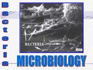
Bacteria • Bacteria are microscopic organisms whose single cells have neither a membrane-enclosed nucleus nor other membrane-enclosed organelles like mitochondria and chloroplasts. • Until recently classification has done on the basis of such traits as:shape • bacilli: rod-shaped • cocci: spherical • spirilla: curved walls • ability to form spores • method of energy production (glycolysis for anaerobes, cellular respiration for aerobes) • nutritional requirements • reaction to the Gram stain. • More recently, genome sequencing, especially of their 16S ribosomal RNA (rRNA), has provided additional insights into the evolutionary relationships among the bacteria.
Pathogenic and Non-pathogenic • Gram negative rods and cocci • Pseudomonads. Pseudomonads are aerobic, Gram-negative rods that are motile with polar flagella. Over 30 species are found in the group, and Pseudomonas fluorescens is a well-known producer of a yellow-green pigment. Another species, P. aeruginosa, causes urinary tract infections and infections of burned tissue.
Enterobacteria. • Over 25 genera of enterobacteria are recognized, many with pathogenic importance. Among the medically important enterobacteria are Salmonella species that cause intestinal disease known as salmonellosis; Yersinia pestis, the cause of plague; Klebsiella species, the causes of pneumonia, intestinal disease, and other infections; and species of Serratia and Proteus. The well-known organism Escherichia coli is also a member of this group. All enterobacteria have peritrichous flagella.
Vibrios • Vibrios are curved, Gram-negative, facultatively anaerobic rods. They belong to the family Vibrionaceae. One species, Vibrio cholerae, is the cause of cholera in humans. Dying of dehydration, Poor sanitation
Sulfur bacteria • use sulfur or sulfur compounds as electron acceptors in their metabolism. These bacteria produce large amounts of hydrogen sulfide during their growth, and therefore, they produce foul odors in water and mud. Members of the genus Desulfovibrio are particularly important in the sulfur cycle for their ability to use sulfur and convert it to other compounds that can be used by plants to synthesize sulfur-containing amino acids.
Streptococci • Streptococci are spherical bacteria that divide in parallel planes to produce chains. The bacteria are Gram-positive, and certain species are aerobic, while others are anaerobic. On blood agar, certain species partly destroy the red blood cells and are said to be alpha-hemolytic. Other species completely destroy the blood cells and are beta-hemolytic. Those streptococci producing no blood cell destruction are gamma-hemolytic.
Staphylococci • Staphylococci are Gram-positive bacteria that divide in planes to produce clusters or packets. Normally associated with the skin and mucous membranes, certain species of staphylococci are involved in skin boils, abscesses, and carbuncles, especially if they produce the enzyme coagulase, which causes blood clotting. Staphylococcus aureus is involved in cases of food poisoning, toxic shock syndrome, pneumonia, and staphylococcal meningitis.
Lactobacilli • Gram-positive, rod-shaped bacteria occurring as single cells or chains. They produce lactic acid in their metabolism and are associated with the flora of the mouth and the vagina. Certain species are associated with the production of dairy products such as yogurt, sour cream, and buttermilk.
Bacillus and Clostridium species • Species of Bacillus and Clostridium are Gram-positive, rod-shaped bacteria able to produce highly resistant endospores (spores). The spores are found in the soil, air, and all environments of the body. Species of Bacillus grow aerobically, and Bacillus anthracis is the cause of anthrax. Clostridium species grow anaerobically, and different species cause tetanus, botulism, and gas gangrene.
Submicroscopic Bacteria EVEN SMALLER BACTERIA
Rickettsiae • Rod-shaped and coccoid bacteria belonging to the order Rickettsiales. These bacteria cannot be seen with the light microscope, and therefore the Gram stain is not used for identification. However, their walls have the characteristics of Gram-negative cell walls. Rickettsiae are obligate intracellular parasites that infect humans as well as arthropods such as ticks, mites, and lice. They are cultivated only with great difficulty in the laboratory and generally do not grow on cell-free media. Tissue cultures and fertilized eggs are used instead
Chlamydiae • Extremely tiny bacteria, below the resolving power of the light microscope. Although the Gram stain is not used for identification, the bacteria have cell walls resembling those in Gram-negative bacteria. • Chlamydiae display a growth cycle that takes place within host cells. The bacteria invade the cells and differentiate into dense bodies called reticulate bodies. The reticulate bodies reproduce and eventually form new chlamydiae in the host cell called elementary bodies.Chlamydiae cause several diseases in humans, such as psittacosis, a disease of the lung tissues; trachoma, a disease of the eye; and chlamydia, an infection of the reproductive tract.
Mycoplasmas • Extremely small bacteria, below the resolving power of the light microscope. They lack cell walls and are surrounded by only an outer plasma membrane. Without the rigid cell wall, the mycoplasmas vary in shape and are said to be pleomorphic. Certain species cause a type of mild pneumonia in humans as well as respiratory tract and urinary tract diseases.
Viruses • Noncellulargenetic elements that use a living cell for their replication and have an extracellular state. Viruses are ultramicroscopic particles containing nucleic acid surrounded by protein, and in some cases, other macromolecular components such as a membranelike envelope.
Viral Structure. • Certain viruses contain ribonucleic acid (RNA), while other viruses have deoxyribonucleic acid (DNA). The nucleic acid portion of the viruses is known as the genome. The nucleic acid may be single-stranded or double-stranded; it may be linear or a closed loop; it may be continuous or occur in segments.
An array of viruses. (a) The helical virus of rabies. (b) The segmented helical virus of influenza. (c) A bacteriophage with an icosahedral head and helical tail. (d) An enveloped icosahedral herpes simplex virus. (e) The unenveloped polio virus. (f) The icosahedral human immunodeficiency virus with spikes on its envelope.
How do we fight Viruses? • Antiviral agents • Acyclovir, is used against herpes viruses because this drug prevents the synthesis of DNA during viral replication. Azidothymidine(AZT) is used for patients with HIV infection because this drug also prevents the synthesis of DNA. Gancicloviris used against cytomegaloviruses, and Amantadineis useful against influenza viruses.
Viral Vaccines • Inactivated viruses (“dead viruses”) are unable to replicate in host cells because of some chemical or physical treatment. The Salk vaccine against polio and the yellow fever vaccine. • Attenuated viruses (“live viruses”) are weakened viruses that replicate at a very slow rate in host cells and generally do not produce any symptoms of disease when inoculated to humans. Attenuated viruses are used in the Sabin polio vaccine and in the vaccines against measles and rubella • Genetic engineering methods - Hepatitis B vaccine
Fungi(singular, fungus) • Together with bacteria, fungi are the major decomposers of organic materials in the soil. They degrade complex organic matter into simple organic and inorganic compounds. In doing so, they help recycle carbon, nitrogen, phosphorous, and other elements for reuse by other organisms. Fungi also cause many plant diseases and several human diseases. • Two major groups of organisms make up the fungi. The filamentous fungi are called molds, while the unicellular fungi are called yeasts. The fungi are classified in the kingdom Fungi in the Whittaker five-kingdom system of classification
Nutrition • Fungi grow best where there is a rich supply of organic matter. Most fungi are saprobic (obtaining nutrients from dead organic matter).
Algae • Eukaryotic organisms that have no roots, stems, or leaves but do have chlorophyll and other pigments for carrying out photosynthesis. Algae can be multicellular or unicellular. • Unicellular algae occur most frequently in water, especially in plankton. Phytoplankton is the population of free-floating microorganisms composed primarily of unicellular algae. In addition, algae may occur in moist soil or on the surface of moist rocks and wood. Algae live with fungi in lichens.