Download

1 / 27
270 likes | 294 Views
Explore the intricate layers and functions of the retina, from phototransduction to visual cycle, macular characteristics, blood supply, vitreous structure, symptoms and signs of disorders, examination techniques, risk factors, and diabetic retinopathy management.

E N D
Retina Prof. Faiz Shakarchi
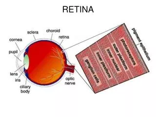
The retina is the photosensitive layer of the eye where light energy is converted to electrical impulses
Retina consists of two main layers: A-The outer layer the Retinal pigment layer (RPE) B-The inner layer the Sensory layer, 1-Photoreceptors (Cones and Rods) 2-Outer limiting membrane 3-Outer nuclear layer 4-Outer plexiform layer 5-Inner nuclear layer 6-Inner plexiform layer 7-Ganglion layer 8-Nerve fiber layer 9-Inner limiting membrane
Phototransduction Visual cycle. Absorption of light by visual pigments (rhodopsin or cone opsin) causes isomerization of 11-cis-retinal to all-trans-retinal. Electrical impulses generated, transmitted to the brain through the optic nerve and the visual pathway.
Macula; an oval area in the posterior pole about 5 mm in diameter, correspond to central 15o of visual field. Fovea; central part in the macula about 1.5 mm in diameter correspond to the central 5o in the visual field. Foveola: central depression in the fovea about 0.35 mm in diameter contains cones only, and correspond to the central 1o of the most precise vision in the visual field.
Oxygen and nutrients supply : Inner layers supplied by central retinal artery Photoreceptors supplied by choriocapillaries
Blood supply to the retina Central retinal artery: Upper and lower temporal arterioles Upper and lower nasal arterioles Retinal vessels are End arterioles
Retina consists of densely packed cells Extra-cellar space is only 1% • Retinal-Blood Barrier: -Inner: tight junctions between the endothelial cells of retinal capillaries - Outer: tight junctions between the retinal pigment epithelial cells
Functions of Retinal pigment epithelium: 1- Regenerates the visual pigments after phototransduction 2- Passage of O2 and nutrients from choroid to the photoreceptors 3- Outer retinal blood barrier 4- Absorb scattered light
The vitreous: a clear gel occupying two-thirds of the globe, consists: - water 98%. - hyaluronic acid - fine collagen network - There are few leukocytes. Vitreous firmly Attached to the peripheral retina, andaround the optic disc.
Symptoms of retinal disorders: 1- Painless impairment of vision. 2 - Distorted vision (metamorphopsia) caused by a disturbance in the arrangement of the photoreceptors in macular diseases such as reduction (micropsia) or enlargement (macropsia) of object size 3-Impairment of color vision which occurs in macular diseases 4- Visual field defects 5-Floaters (perception of moving images in the field of vision, caused by vitreous opacities that cast a shadow on the retina). 6-Photopsia (perception of flashes of light)
Signs 1-Depressed Visual acuity 2-Impairment of Pupillary light reflex 3-Vitreous opacities Hemorrhage WBC Pigment dots (Tobacco dust) 4-Retinal hemorrhage - Hard exudates: yellow spots well demarcated margins , deposition of lipoproteins, or lipid, are sign s of abnormal vascular leakage - Cotton wool spots: fluffy white spots with indistinct margins, accumulation of axoplasmic debris in the nerve fiber layer , they are sins of retinal ischemia (micro-infraction of the nerve fiber layer) 5-Abnormal position (Retinal detachment) 6-Neo-vascularization: retinal ischemia ; secretion of vaso-formative factors NVD (neo-vascularization on the surface of the optic disc) NVE (neo-vascularization on the surface of the retina).
Examination of the retina • Direct ophthalmoscope • Indirect ophthalmoscope Investigations • Fluorescein angiography-FA • Optical coherence tomography-OCT
Risk factors: 1-Duration of diabetes. After 10 years 50% have retinopathy, while after 30 years 90% have retinopathy 2-Poor metabolic control 3-Hypertension 4-Nephropathy 5-Pregnancy 6-Others; smoking, obesity, hyperlipidaema. Diabetic RetinopathyOne of the most important causes of blindness
1- Micro-vascular leakage -microaneurysms -Hard exudates 2- Micro-vascular occlusion Cotton wool spots Formation of abnormal neo-vasculartization on the surface of the retina (NVE, on the optic disc (NVD). and on the Iris (Rubeosis) Pathogenesis:It is a microangiopathy, affecting pre-capillary arterioles, capillaries, and post-capillary venules.
Classification of diabetic retinopathy; 1-Background (non-proliferative) microaneurysms, small Retinal hemorrhages (blot and dots ), and hard exudates.
2-Maculopathy, (clinical significant macular edema). Microaneurysms, hemorrhages, and hard exudates at the macula. • Vision is impaired
3-Pre-proliferative. Large retinal hemorrhage, cotton wool spots (infarction in the nerve fiber layer), venous congestion and dilatation.
4-Proliferative retinopathy. Abnormal neo-vasculartization on the surface of the retina (NVE) and on the optic disc (NVD).
5-Advanced diabetic retinopathy. Vitreous hemorrhage and tractional retinal detachment.
Management: Essential Important Point is: Early Detection of Diabetic Retinopathy The treatment is more effective and the prognosis is better in early stages. Every diabetic patient must has regular ophthalmic examination for detection retinopathy. Management:
Background; good diabetic control Control of other risk factors
Maculopathy: Laser phototherapy. Laser burns are directed at the sites of leakage ( micro-aneurysms), avoiding the central fovea. • Intra-vitreal injection of Anti-vascular endothelial growth factor (Anti-VGEF)
Pre-proliferative and Proliferative retinopathy; Laser phototherapy. The entire retina is treated with laser burns except the macula and area adjacent to the optic disc Pan retinal photocoagulation (PRP). The Laser burns destroy the ischemic retina and prevent release of vaso-formative factors and causing regression of the abnormal vessels.
Advanced retinopathy; Surgery (Pars Plana Vitrectomy). Removal of the vitreous hemorrhage, vitro-retinal bands and endo-laser through small incisions at pars plana (posterior part of the ciliary body).