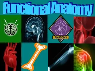
Functional Anatomy
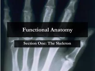
Functional Anatomy. Section One: The Skeleton. Functions of a Skeleton. The skeleton performs 5 basic functions:. 1.3 Axial and Appendicular Skeleton. All the bones of the skeleton are divided into two main groups. These are known as: Axial Skeleton Appendicular Skeleton AXIAL SKELETON

Functional Anatomy
E N D
Presentation Transcript
Functional Anatomy Section One: The Skeleton
Functions of a Skeleton The skeleton performs 5 basic functions:
1.3 Axial and Appendicular Skeleton All the bones of the skeleton are divided into two main groups. These are known as: • Axial Skeleton • Appendicular Skeleton AXIAL SKELETON Consists of those bones forming the central column of the body, i.e. spine, skull and ribcage. APPENDICULAR SKELETON Those bones that attach to the axial skeleton, i.e. shoulders, hips and the limbs.
1.4 Classification of Bones Bones are classified according to their shape. They fall into four basic categories: • Long bones • Short bones • Irregular bones • Flat bones Using the table, fill in the basic function of each type of bone and provide some examples of these.
On the skeleton, colour the short, flat, longand irregularbones you can identify.
What do you notice about the location of most of the flat bones? Why might this be? Located around the main organs, e.g. brain, heart. To give protection. What do you notice about the location of most of the long bones? Why might this be? Located in legs and arms. These are the regions of most joints and therefore movement.
1.5 Identifying Bones of the Skeleton Cranium (skull) As a pre-test, try naming as many bones as possible on the skeleton below. Use common or anatomical terms. Vertebrae – cervical (neck) Sternum (breastbone) Ribs (ribcage) Vertebrae – lumbar (lower back) Pelvis (hip) = Ilium, Ischium & Pubis Metacarpals (palm) Phalanges (fingers) Tibia (shin)
Maxilla (face) Mandible (jaw) Scapula (shoulder blade) Clavicle (collarbone) Ulna (forearm) Humerus (upper arm) Radius (forearm) Femur (thigh) Fibula (shin) Patella (knee cap) Tarsal (heel) Metatarsals (foot) Phalanges (toes)
A. Cranium (common name = skull) Designed to protect the brain. Made up of a number of inter-connecting bones. • THE HEAD Identify and explain the function of the following skeletal structures. A B. Maxilla (common name = face) Houses the eyes and sinuses. Protects these features against damage. B C C. Mandible (common name = jaw) Responsible for talking, chewing etc.
2. THE RIBCAGE Ribs (common name = ribs) 12 pairs in all. Designed to protect the heart and lungs Rib 3. THE CHEST A. Sternum (common name = breast bone ) Protects the heart and lungs. It is the bone pressed on in C.P.R.
Cervical vertebrae. These are small delicate bones responsible for neck movement. There are seven bones in all. 4. THE SPINE Thoracic vertebrae. Allow ribs to attach to the spine hence there are 12 of them (one for each rib pair). Lumbar vertebrae. These are the largest of the vertebrae and are responsible for weight-bearing. There are five in all. The sacrum (upper) and coccyx (lower) are a series of fused (joined) bones that help form the pelvis.
What do the shapes of the bones tell you about their function? Those that are larger have a roll in weight or load bearing e.g. lumbar and thoracic. Smaller ones are important for movement e.g. cervical. 5. THE SHOULDER A. Scapula (common name = shoulder blade) Protects the lungs. Forms shoulder joint. B. Clavicle (common name = collarbone) Holds the shoulder in place. Easily broken. B A
A. Humerus (common name = upper arm ) Prime function is movement. B. Radius (common name = Forearm ) Prime function is movement, always located on thumb-side of forearm. C. Ulna (common name = forearm ) Prime function is movement. D. Carpels (common name = wrist ) Prime function is movement. E. Metacarpels (common name = palms ) Prime function is movement. F. Phalanges (common name = Fingers ) Prime function is movement. 6. THE ARM A B C D E F
A. Ilium (common name = pelvis) Protects intestines B. Pubis ( common name = pelvis) Forms front of pelvis. Has to separate in childbirth. C. Ischium (common name = pelvis) Forms the ‘boney bum’. A 7. THE PELVIS B C
A. Femur (common name = thigh ) Largest bone in the body, responsible for support and movement. B. Patella (common name = knee cap ) Protects the knee joint. C. Tibia (common name = shin ) Support and movement. D. Fibula (common name = shin) ‘Thinner’ bone of leg. Support and movement. E. Tarsels (common name = ankle) Bones of the ankle and heel. Support and balance. F. Metatarsels (common name = foot) Form the sole of the foot. Support and balance. G. Phalanges (common name = toes) Support, movement and balance. A B C D E F G 8. THE LEG
Functional Anatomy Section Two: Terms of Direction
2.2 The Anatomical Position In order to explain the positioning of bones, organs, muscles and the like on the human body, anatomists have agreed on a standardised position for the human body in all cases. This is known as the anatomical position. THE ANATOMICAL POSITION There are four key features to note: • Palms face forward • Body is upright • Thumbs point outward – so radius and ulna and uncrossed • Face is forward
The terms of direction in the next section are all with respect to this position. Why is it important to always talk about the position of organs, bones and muscles in or on the human body with respect to the anatomical position? This enables everyone to talk from the same point of view regardless of their profession or level of expertise.
2.3 Anatomical Terms of Direction These refer to the position of parts of the body, or of one part with respect to another.
Iliopsoas Pectoralis Major This exercise has shown that the terms of direction compliment each other. Complete the list below by placing the opposite term next to the one provided. Superior - InferiorAnterior - Posterior Proximal - Distal Medial - Lateral Deep - Superficial Supine - Prone
Functional Anatomy Section Three: The Joints
3.2. Overview of Joint Types There are three broad categories of joint type in the body. They are classed according to the degree of movement possible. The three categories are: 1. Immovable Also known as fibrousjoints 2. Slightly movable Also known as cartilaginousjoints 3. Freely movable Also known as synovialjoints We shall look at all these categories in turn.
3.3 Fibrous Joints These are non-movable joints. They are the result of tough fibrous tissue forming where the two bone ends meet. What is the function of a fibrous joint? To provide protection. Examples include: 1. Skull 2. Pelvis Fibrous joint
3.4 Cartilaginous Joint These are slightly-movable joints. They are the result of cartilage forming where the two bone meet. This gives a fair degree of resilience. What is the function of a cartilaginous joint? To act as shock absorbers. Examples include: 1. Invertebral discs 2. Ribs to sternum 3.Where pubic bones meet Cartilaginous Joints
3.5 Synovial Joint These are freely movable joints. The only limitation in range of movement is as a result of bone shape at the joint, and ligaments. What is the primary function of a synovial joint? To provide movement. All synovial joints follow the same basic structure as shown
The key components of your illustration have important roles to play in maintaining the structure of the joint. • Ligaments Join bone to bone for stability 2. Capsule Provides stability and protection from infection 3. Cartilage Reduce wear and tear on bones 4. Synovial Fluid Lubricates the joint and provides shock absorption 5. Synovial Membrane Produces synovial fluid In some joints, for example the knee, there are pads of fat and/or discs of cartilage to further help absorb shock and reduce general ‘wear and tear’.
3.6 Types of Synovial Joints Synovial joints can be divided into six basic types. The types are governed by the type of movement or movements they allow. The six basic types are: • Gliding • Hinge • Pivot • Condyloid • Saddle • Ball and Socket
1. Gliding Definition: The bone surfaces are small and flat, or slightly concave and one bones slides over the other. Examples: 1. Carpals and tarsals 2. Ribs and vertebrae 3. Scapula and ribs Movements: Only slight movement is possible due to the restrictions of attached ligaments. Movements possible are: 1. side to side (abduction / adduction) 2. Back and forth (extension/flexion)
2. Hinge Definition: Two bones join in such a way that movement is possible only in one direction, usually at right angles to the bones. Examples: 1. Elbow 2. Knee 3. Ankle Movements: A uniaxial joint allowing movement in only one direction The only movement possible is: Back and forth (extension/flexion)
3. Pivot Definition: A joint constructed in such a way that rotation only is possible (usually about the long axis of the bone) Examples: 1. Atlas and axis of neck 2. Radius and humerous Movements: A uniaxial joint allowing movement in only one direction The only movement possible is: Rotation
4. Condyloid Definition: Also known as an ellipsoid joint. The bone ends make the shape of an ellipse. Examples: 1. Carpals and radius 2. Metacarpals and phalange Movements: A biaxial joint allowing movement in two main directions. The movements possible are: 1. Back and forth (extension/flexion) 2. Side to side (abduction/adduction) 3. Some Circumduction
5. Saddle Definition: The bone ends are shaped like a rider on a saddle Example: 1. Carpal/metacarpal of thumb Movements: A biaxial joint allowing movement in two main directions. Movements possible are: 1. side to side (abduction / adduction) 2. Back and forth (extension/flexion)
6. Ball and Socket Definition: A ball-shaped bone end fits into a socket or cup-shaped bone. Examples: 1. Hip 2. Shoulder Movements: A multiaxial joint allowing movement in many directions around the joint. The movements possible are: 1. Back and forth (extension/flexion) 2. Side to side (abduction/adduction) 3. Rotation 4. Circumduction
The shoulder joint is the most freely moving ball and socket joint we have. The illustration may help you with your answer. Why is the shoulder joint so freely moving? Because the socket is shallow. What do you suppose is the risk of such a freely moving joint? It is easy to dislocate.
3.7. Movements at Synovial Joints Just as we learnt a set of terms to describe the positioning of bones, muscles and organs in the body, so we have a set of terms to describe how joints move.
Functional Anatomy Section Four: The Muscles
4.2 Identifying Muscles Trapezius Deltoid Biceps Triceps Pectoralis Major Latissimus Dorsi Rectus Abdominus Gluteus Maximus Hamstrings Quadriceps Gastrocnemius
Rectus Abdominus 4.3 Guide to Individual Muscles [A] PRIME MOVERS OF THE TRUNK • Rectus adbominus: Location : A group of two muscles running lengthwise along the medial aspect of the abdomen. They are rather like two columns of muscle running up either side of the belly button. They run from the pubis (pelvis) to the cartilage of the 5th, 6th and 7th ribs. Movements: Rectus abdominus allows two basic movements. 1. Flexion of the trunk 2. Lateral flexion of the trunk Application: Typical sporting actions include: 1. Sit ups 2. Cartwheels 3. Pike in diving
Erector Spinae [A] PRIME MOVERS OF THE TRUNK 2. Erector Spinae Group: Location : One of the main muscles located in the lower back and one of the few visible. It runs from the pelvis to the lumbar vertebrae. Movements: Erector spinae group allows two basic movements. They are: 1. Extension of the spine 2. Lateral flexion of the trunk Application Typical sporting actions include: 1. Straightening out from a pike 2. Rowing 3. Swimming (body position)
Trapezius [B] PRIME MOVERS OF THE SHOULDER 1. Trapezius: Location : A large triangular muscle located on the posterior aspect of the body. It runs in a triangular shape from the base of the skull, the 7th cervical vertebrae and all the thoracic vertebrae, to the scapula and clavicle. Movements: Trapezius allows four basic movements. They are: • Raise the head • Pull the shoulders back • Raise the scapula 4. Drop the scapula Application: Typical sporting actions include: 1.Pulling shoulder when throwing 2. Rowing 3. Looking up in basketball
Latissimus dorsi [B]PRIME MOVERS OF THE SHOULDER 2. Latissimus Dorsi: Location : The broadest muscle of the back. It forms the back of the armpit. This is a large triangular muscle which covers the lumbar and lower thoracic region of the back. It runs from the lower thoracic and lumber regions, to the anterior aspect of the humerus Movements: Latissimus dorsi allows three basic movements. They are: 1. Adduction of the upper arm 2. Extension of the shoulder 3. Internal rotation of the shoulder How can latissimus dorsi allow internal rotation of the shoulder to occur if it is a muscle located on the back? Because it attaches to the humerus Application: Typical sporting actions include: 1. Recovery in breaststroke 2. Ten-pin bowling 3. Drawing arm back to punch
Deltoid [B]PRIME MOVERS OF THE SHOULDER 3. Deltoid: Location : A triangular shaped muscle located on the superior aspect of the shoulder i.e. above the shoulder joint It runs from the scapular and clavicle to attach at the humerus Movements: Deltoid allows four basic movements. They are: 1. Flexion of the shoulder 2. Extension of the shoulder 3. Abduction of the arm 4. Rotation of the shoulder Application: Typical sporting actions include: 1. Throwing 2. Punching 3. Swimming
Pectoralis major [B]PRIME MOVERS OF THE SHOULDER 3. Pectoralis Major: Location : A large triangular muscle located on the chest. It runs from the clavicle, sternum and the 6th rib, to the humerus. Movements: Pectoralis major allows three basic movements. These are: 1. Flexion of the shoulder 2. Abduction of the arm 3. Rotation (internal) of the shoulder Application: Typical sporting actions include: 1. Throwing 2. Punching 3. Press-ups