Download
1 / 33
340 likes | 420 Views
Dive into the various disorders of the esophagus including motility issues, diverticula, and esophagitis. Learn about symptoms, diagnostic methods, and treatment options for each condition.

E N D
Pathology of the Esophagus Christopher Bibb, M.D. Brazos Valley Pathology
Obstruction due to motility disorders • Nutcracker esophagus: Peristaltic pressures exceeding 180 mmHg in the lower esophagus • Diffuse esophageal spasms: Uncoordinated contractions of the esophagus • Presentation: Dysphagia and chest pain • Both conditions are non-progressive • Symptomatic treatment: nitrates or calcium-channel blockers • Can cause small diverticula “Corkscrew esophagus”
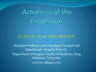
Achalasia – 1 • Infrequent esophageal disorder (1.6 cases/100,000) in which the lower esophageal sphincter (LES) fails to relax, blocking the passage of food bolus • Proximal esophagus dilates progressively • Symptoms: Dysphagia, regurgitation, chest pain, weight loss Barium swallow shows failure of esophagus to empty at 5 minutes. Note bird’s beakappearance of LES
Achalasia – 2 • Idiopathic: Most frequent, due to loss of nitrergic (NO) inhibitory neurons in the myoenteric plexus • Secondary:Chagas disease (Trypanosomacruzi), diabetic autonomic neuropathy, cancer, amyloidosis, sarcoidosis
Zenkerdiverticulum – 1 Killian triangle in the posterolateral aspect of upper esophagus: Area of wall weakness where Zenker diverticulum develops • Impaired relaxation of CPM, which acts as the upper esophageal sphincter, leading to excessive intraluminal pressure in the distal pharynx • Outpouching of mucosajust above the CPM • Pulsion diverticulum, as opposed to traction diverticulum
Zenkerdiverticulum – 2 • Symptoms:Dysphagia, sense of lump in the neck, regurgitation, cough, halitosis • Diagnosis: Made by X-rays after barium swallow • Endoscopy: Risk of perforation • Surgical resection or endoscopic stapling for symptomatic diverticula Barium swallow: Collection of barium within diverticulum located posteriorly, at midline, just above the CPM. It protrudes laterally (usually to the left), and caudally.
Esophageal Webs • Ledge-like mucosal folds of unclear origin • More frequent in women in their 40s • Manifest with dysphagia, food impaction • Associated with GERD, bullous diseases of skin, graft-versus-host disease • Plummer-Vinson syndrome: Esophageal webs, iron-deficiency anemia, glossitis and cheilosis, increased risk of squamous cell carcinoma
Esophageal Rings – Schatzki rings • Circumferential rings of esophageal mucosa/submucosa • Schatzki A rings: above GE junction, not fixed, infrequent • Schatzki B rings: at GE junction, fixed, frequent • Present in 6% of population • May manifest with dysphagia, food impaction Schatzki B ring (arrows) visualized on top of a sliding hiatal hernia (arrowhead)
Mallory-Weiss tears • Mucosal tears at the GE junction • Caused by prolonged or forceful retching, vomiting or coughing • Alcohol abuse and eating disorders predisposing conditions • Hematemesis or melena • Bleeding stops spontaneously • Treatment rarely necessary: cauterization or epinephrin injection • Boerhaave syndrome: Vomiting-induced esophageal rupture described in a drunken Dutch admiral Mallory-Weiss tears From Damjanov and Linder
Esophagitis • Chemical/physical: Alcohol, smoking, hot food, pill stuck to the mucosa (pill esophagitis), radiation, chemotherapy • Usually mild and self-limited • Manifests with painful dysphagia • Rarely severe, may lead to scarring and strictures • Infectious: Usually affects immune depressed patients • HSV, CMV and Candida most frequent causes
Infectious esophagitis Herpes nuclear inclusions CMV nuclear and cytoplasmic inclusions Viral esophagitis: red, ulcerated mucosa in distal esophagus Candidaesophagitis: Gray-white pseudomembranescomposed of organisms and inflammatory debris
Eosinophilic esophagitis – 1 • Most frequent in children • Presentation: Heartburn, dysphagia, food impaction, food intolerance • Lack of response to PPI treatment
Eosinophilic esophagitis – 2 • Diagnosis: Endoscopy and biopsy • Infiltration of epithelium by eosinophils • Proximal esophagus preferentially involved • Pathogenesis: Probably due to food allergy • Treatment: Elimination of allergen, oral corticosteroids Numerous intraepithelial eosinophils with eosinophilic microabscesses.
Reflux esophagitis – 1 • Gastro-Esophageal Reflux Disease (GERD): The most common cause of esophagitis • Protective factors: • Squamous stratified epithelium • Mucin production by submucosal glands • Tone of LES • Predisposing factors:Alcohol, smoking, obesity, pregnancy, CNS depressants, hiatal hernia Normal esophageal mucosa
Reflux esophagitis – 2 Severe reflux esophagitis. Hyperemia with multifocal ulceration. Histology: Ulceration and inflammation of the mucosa, associated with epithelial hyperplasia.
Reflux esophagitis – 3 • Frequent symptoms: Heartburn, occasional regurgitation, dysphagia • Less frequent symptoms: Angina-like pain, odynophagia, increased salivation, nausea • Atypical symptoms: Chronic cough, laryngitis with hoarseness, asthma, erosion of enamel, possibly pulmonary fibrosis, sinusitis and otitis • Complications: • Strictures due to ulcers and scarring • Barrett esophagus and adenocarcinoma • Treatment: Proton pump inhibitors (PPIs)
Barrett esophagus Intestinal metaplasia of the esophageal epithelium in the lower esophagus. Stratified squamous epithelium is replaced by intestinal epithelium with goblet cells. Barrett esophagus: 10% of patients with GERD Dysplasia:0.2% -2% of patients with Barrett
Barrett: Gross changes Endoscopic appearance of Barrett esophagus Barrett esophagus (red mucosa above GE junction) Normal gastroesophageal junction GE junction
Barrett: Microscopic changes Barrett: Intestinal type epithelium with hgh grade dysplasia. Note architectural disorganization, high nucleo-cytoplasmic ratio and absence of goblet cells. Barrett: Intestinal type epithelium. Note globlet cells. No dysplasia.
Barrett esophagus: Premalignant condition • One in 200 patients with Barrett esophagus will develop cancer every year • Management: • No dysplasia or low-grade dysplasia: Endoscopic monitoring • High grade dysplasia or intramucosal carcinoma:Esophagectomy or other surgical methods of resection (endoscopic mucosal resection, laser ablation) Norman Barrett (1903-1979) From: Eponyms in Radiology of the Digestive Tract: Historical Perspectives and Imaging Appearances Radiographics, 2006, 26: 129 By J.P. Kanne et al.
Chronic gastroesophageal reflux Barrett esophagus Low-grade dysplasia High-grade dysplasia Adenocarcinoma
Esophageal adenocarcinoma:Changing epidemiology • At present, adenocarcinomas outnumber SCCs • From the 70s to now incidence rose more rapidly than any other cancer • M:F ratio = 7:1 • Caucasian men are more affected • Main risk factors: • GERD and Barrett esophagus • Obesity:FOURfold increase • Smoking • Alcohol, but less than squamous cell carcinoma • H. pylori possibly protective due to decreased reflux
Esophageal Adenocarcinoma Endoscopic view of esophageal adenocarcinoma: almost always in the distal third Adenocarcinoma (right) in Barrett mucosa (left)
Esophageal Adenocarcinoma: Manifestations • Progressive dysphagia • Weight loss • Chest pain • Hoarseness, late sign, infiltration of recurrent laryngeal nerve • Aspiration pneumonia, late sign
Esophageal Adenocarcinoma: Prognosis • Spread to mediastinal organs, regional lymph nodes and distant organs • Stage is the most important prognostic factor • Overall 5-year survival rate: < 25% • Limited to mucosa-submucosa: 80% Overall 5-yr survival of 402 patients with esophageal adenocarcinoma
Esophageal Squamous Cell Carcinoma (SCC) • Risk factors: • Smoking and alcohol • Poverty • Achalasia, radiation, caustic injury, Plummer-Vinson syndrome, hot beverages • M:F ratio: 4:1 • B:C ratio: 6:1 • Begins as squamous dysplasia • Clinical presentation similar to adenocarcinoma NORMAL DYSPLASIA
Esophageal SCC SCC occurs most often in the middle-third Often causes strictures Composed of disorganized squamous epithelium
Esophageal SCC: Prognosis • Treatment: Combination of surgery, radiation, chemotherapy • Stage based outcome: < 25% survive at 5 years despite all advances in surgery, radiation therapy and chemotherapy • Mucosal disease 80% 5 year survival • Palliation is often the only option