Download
1 / 36
410 likes | 807 Views
Chapter 7 The Nervous System: Structure and Control of Movement. EXERCISE PHYSIOLOGY Theory and Application to Fitness and Performance, 6th edition Scott K. Powers & Edward T. Howley. General Nervous System Functions. Control of the internal environment With the endocrine system

E N D
Chapter 7The Nervous System: Structure and Control of Movement EXERCISE PHYSIOLOGY Theory and Application to Fitness and Performance, 6th edition Scott K. Powers & Edward T. Howley
General Nervous System Functions • Control of the internal environment • With the endocrine system • Voluntary control of movement • Programming spinal cord reflexes • Assimilation of experiences necessary for memory and learning
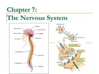
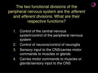
Organization of the Nervous System • Central nervous system (CNS) • Brain and spinal cord • Peripheral nervous system (PNS) • Neurons outside the CNS • Sensory division • Afferent fibers transmit impulses from receptors to CNS • Motor division • Efferent fibers transmit impulses from CNS to effector organs
Anatomical Divisions of the Nervous System Figure 7.1
Relationship Between PNS and CNS Figure 7.2
Structure of a Neuron • Cell body • Dendrites • Conduct impulses toward cell body • Axon • Carries electrical impulse away from cell body • May be covered by Schwann cells • Forms discontinuous myelin sheath along length of axon • Synapse • Contact points between axon of one neuron and dendrite of another neuron
The Parts of a Neuron Figure 7.3
Synaptic Transmission Figure 7.4
Electrical Activity in Neurons • Neurons are “excitable tissue” • Irritability • Ability to respond to a stimulus and convert it to a neural impulse • Conductivity • Transmission of the impulse along the axon
Resting Membrane Potential • Negative charge inside cells at rest • -5 to -100 mv (-40 to -75 mv in neurons) • Determined by: • Permeability of plasma membrane to ions • Difference in ion concentrations across membrane • Na+, K+, Cl-, and Ca+2 • Maintained by sodium-potassium pump • Potassium tends to diffuse out of cell • Na+/K+ pump moves 2 K+ in and 3 Na+ out
The Resting Membrane Potential in Cells Figure 7.5
Illustration of Ion Channels Figure 7.7
The Sodium-Potassium Pump Figure 7.8
Action Potential • Occurs when a stimulus of sufficient strength depolarizes the cell • Opens Na+ channels and Na+ diffuses into cell • Inside becomes more positive • Repolarization • Return to resting membrane potential • K+ leaves the cell rapidly • Na+ channels close • All-or-none law • Once a nerve impulse is initiated it will travel the length of the neuron
An Action Potential Figure 7.9
Depolarization and Repolarization of a Nerve Fiber Figure 7.10
Neurotransmitters and Synaptic Transmission • Synapse • Small gap between presynaptic neuron and postsynaptic neuron • Neurotransmitter • Chemical messenger released from presynaptic membrane • Binds to receptor on postsynaptic membrane • Causes depolarization of postsynaptic membrane
Basic Structure of a Chemical Synapse Figure 7.11
Neurotransmitters and Synaptic Transmission • Excitatory postsynaptic potentials (EPSP) • Causes depolarization • Temporal summation • Summing several EPSPs from one presynaptic neuron • Spatial summation • Summing from several different presynaptic neurons • Inhibitory postsynaptic potentials (IPSP) • Causes hyperpolarization
Proprioceptors • Provide CNS with information about body position and joint angle • Free nerve endings • Sensitive to touch and pressure • Initially strongly stimulated then adapt • Golgi-type receptors • Found in ligaments and joints • Similar to free nerve endings • Pacinian corpuscles • In tissues around joints • Detect rate of joint rotation
Muscle Chemoreceptors • Sensitive to changes in the chemical environment surrounding a muscle • H+ ions, CO2, and K+ • Provide CNS about metabolic rate of muscular activity • Important in regulation of cardiovascular and pulmonary responses
Reflexes • Rapid, unconscious means of reacting to stimuli • Order of events: • Sensory nerve sends impulse to spinal column • Interneurons activate motor neurons • Motor neurons control movement of muscles • Reciprocal inhibition • EPSPs to muscles to withdraw from stimulus • IPSPs to antagonistic muscles • Crossed-extensor reflex • Opposite limb supports body during withdrawal of injured limb
The Crossed-Extensor Reflex Figure 7.12
Somatic Motor Function • Somatic motor neurons of PNS • Responsible for carrying neural messages from spinal cord to skeletal muscles • Motor unit • Motor neuron and all the muscle fibers it innervates • Innervation ratio • Number of muscle fibers per motor neuron
Illustration of a Motor Unit Figure 7.13
Vestibular Apparatus and Equilibrium • Vestibular apparatus • Located in the inner ear • Responsible for maintaining general equilibrium and balance • Sensitive to changes in linear and angular acceleration
Role of the Vestibular Apparatus in Maintaining Equilibrium and Balance Figure 7.14
Motor Control Functions of the Brain • Brain stem • Responsible for: • Many metabolic functions • Cardiorespiratory control • Complex reflexes • Major structures: • Medulla • Pons • Midbrain • Reticular formation
Motor Control Functions of the Brain • Cerebrum • Cerebral cortex • Organization of complex movement • Storage of learned experiences • Reception of sensory information • Motor cortex • Motor control and voluntary movement • Cerebellum • Coordinates and monitors complex movement
Motor Functions of the Spinal Cord • Withdrawal reflex • Other reflexes • Important for the control of voluntary movement • Spinal tuning • Voluntary movement translated into appropriate muscle action
Control of Motor Function • Subcortical and cortical motivation areas • Sends a “rough draft” of the movement • Cerebellum and basal ganglia • Coverts “rough draft” into movement plan • Cerebellum: fast movements • Basal ganglia: slow, deliberate movements • Motor cortex through thalamus • Forwards message sent down spinal neurons for “Spinal tuning” and onto muscles • Feedback from muscle receptors and proprioceptors allows fine-tuning of motor program
Structures and Processes Leading to Voluntary Movement Figure 7.16
Autonomic Nervous System • Responsible for maintaining internal environment • Effector organs not under voluntary control • Smooth muscle, cardiac muscle, and glands • Sympathetic division • Releases norepinephrine (NE) • Excites an effector organ • Parasympathetic division • Releases acetylcholine (ACh) • Inhibits effector organ
Neurotransmitters of the Autonomic Nervous System Figure 7.17
Exercise Enhance Brain Health • Exercise improves brain function and reduces the risk of cognitive impairment associated with aging • Regular exercise can protect the brain against disease (e.g. Alzheimer’s) and certain types of brain injury (e.g. stroke)