Download
1 / 54
560 likes | 760 Views
Iliac Disease: Core Curriculum. Iliac Disease. Diagnosis Indications Technical Issues Treatment Options - PTA - Surgical Complications Prognosis. Iliac Disease : Initial Assessment. Physical examination signs of peripheral ischemia

E N D
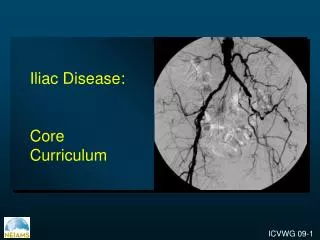
Iliac Disease: Core Curriculum
Iliac Disease Diagnosis Indications Technical Issues Treatment Options - PTA - Surgical Complications Prognosis
Iliac Disease : Initial Assessment • Physical examination • signs of peripheral ischemia • distal embolization • status of the peripheral pulses. • Rest and exercise ABI
Iliac Disease: Diagnosis Noninvasive imaging modalities • Pressure Gradients • Duplex ultrasound (DUS) scans • Magnetic resonance angiography (MRA) • Computed tomography angiography (CTA)
Pressure Gradients obtained during revascularization of iliac occlusion A. Baseline gradient. B. Gradient after administration of nitroglycerine. C. Postballoon, significant resting gradient remains, evn without provocation. D. Gradient eliminated after stenting, demonstrating superior hemodynamic result. Grossmans “Catheterization” 7th Ed. pg. 588-592.
Iliac Disease: Duplex Ultrasound • DUS has proved to be cost-effective and accurate for the detection of significant vascular stenoses and is therefore often used as the first diagnostic modality. 1, 2 • The poor monophasic duplex waveform at the common femoral artery is in itself an accurate marker of aortoiliac obstructive disease. Other waveforms are nondiagnostic for aortoiliac disease.3 • Kohler et al Ann vasc surg 1990 (4) 280-287 • Visser et al Radiology 2000 (16) 67-77 • Spronk et al J vasc surg 2005; 42(2): 236-242
Triphasic Biphasic : Three waveform “phases” consisting of a sharp systolic forward up rise and fall, an element of reverse flow during diastole, and an element of forward flow during diastole Two waveform “phases” consisting of a sharp systolic forward up rise and fall and an element of reverse flow during diastole Sharp Monophasic Poor (blunted) monophasic One waveform “phase” with a sharp systolic rise, the lack of a reverse diastolic element, and a fast diastolic fall, expected in arterial segments proximal to an obstruction The loss of “sharpness” in systole, the lack of a reverse diastolic element, and a slow diastolic fall expected in arterial segments distal to an obstruction Spronk et al J vasc surg 2005; 42(2): 236-242
Iliac Disease: MR Angiography • Enhanced MR angiography showed significant improvement (P < .001) compared with unenhanced MR angiography for diagnosis of clinically significant aortoiliac occlusive disease Rapp et al Radiology 2005; 236: 71-78
Iliac Disease: MR Angiography Gadofosveset-enhanced MR angiography Transverse reconstruction of a steady-state gadofosveset dataset showing stenoses (arrows) in both right and left common iliac arteries. Rapp et al Radiology 2005; 236: 71-78
Iliac Disease: Computed Tomography Angiography • CT angiographic examination is less invasive and less expensive than conventional angiography • Improves resolution with decreased contrast load and acquisition time without increasing radiation exposure A 4-channel MDCT angiogram: Coronal curved planar reformatted images of the abdominal aorta and right iliac artery Rubin et al 2000 Radiology 215: 63-70 Karcaaltincaba M, Foley D Cardiovasc Interv Rad2005; 28(2): 169-172
Morphological Stratification of Iliac Lesions ACC/AHA Guidelines
Morphological Stratification of Iliac Lesions ACC/AHA Guidelines
Iliac Disease Diagnosis Indications Technical Issues Treatment Options - PTA - Surgical Complications Prognosis
Indications for Revascularization • Relief of symptomatic lower extremity ischemia, including claudication, rest pain, ulceration or gangrene, or embolization causing blue toe syndrome www.emedicine.com • Restoration y/o preservation of inflow to the lower extremity in the setting of pre-existing or anticipated distal bypass • Procurement of access to more proximal vascular beds for anticipated invasive procedures. Occasionally revascularization is indicated to rescue flow-limiting dissection complicating access for other invasive procedures Grossmans “Catheterization” 7th Ed. pg. 588-592.
Specific Indications for Revascularization Iliac artery revascularization before cardiac surgery • Significant bilateral disease in order to allow the intra-aortic balloon pump insertion Rigateli et al Internat J Cardiovasc Imag 2002; 22:305-310
Iliac Disease Diagnosis Indications Technical Issues Treatment Options - PTA - Surgical Complications Prognosis
Diagnostic aortogram: Inflow and outflow of the target lesion Run-off angiography: Visualization of the lower extremity circulation Iliac Disease: Angiography Transbrachial aortography documents a TransAtlantic Inter-Society Consensus class D iliac occlusion with right external iliac occlusion and complete occlusion of the left iliac system in a 46-year-old man with disabling claudication Leville et al J Vasc Surg 2006; 43(1):32-39
Iliac Disease: Technical Issues • Endovascular Access • Ipsilateral femoral artery • Contralateral femoral artery • Brachial artery: In patients with flush occlusions at the aortic bifurcation • Multiple access sites may be required for successful treatment: • Bilateral femoral • Femoral/brachial Endovascular recanalization was performed with a hydrophilic guidewire and catheter, and femoral access was obtained with ultrasound guidance Leville et al J Vasc Surg 2006; 43(1):32-39
Iliac Disease: Technical Issues Anticoagulation • Aspirin (325 mg) once a day several days prior to the procedure • Heparin (2500-5000 IU) after access has been obtained and prior to the intervention
Iliac Disease Diagnosis Indications Technical Issues Treatment Options - PTA - Surgical Complications Prognosis
Iliac Artery Disease: Treatment Percutaneous transluminal angioplasty (PTA) with or without implantation of a stent is still considered as the gold standard in the treatment of a peripheral lesion.
Interventional Management of Iliac Lesions • Endovascular treatment of iliac stenoses • High technical success rates • Low morbidity • Iliac PTA/stenting • High rates of patency • Improvement in functional outcome for the individual patient Bosch et al Circulation 1999; 99:3155-3160
Iliac Artery Disease: PTA Iliac Angioplasty: metaanalysis of 2697 procedures before 1990 75% claudicants 2 year primary patency of 81% 5 year primary patency rate 75% Becker, Radiology 1989 Short segment Iliac stenoses: PTA has 5 year 80-90% patency rate Pentacost Circulation 1994
Iliac Artery Disease: Surgical vs PTA Randomized Data • VA Randomized Study: • Patients with limited disease suitable for PTA or surgery. • Excluding initial PTA failure rate of 15%, 3- year • patency of 75% was equivalent in both arms Wilson SE, J Vasc Surg 1989 • Swedish Study: • Equivalent 1 year results Holm, Eur J Vasc Surgery 1991
Iliac Artery Disease: Stent vs PTA • Kauffmann 1991: BE stent vs PTA • Randomized trial enrolled 131 patients • 2 year clinical patency: • 89% after stent, • 70% after PTA. • Bosch 1997: meta-analysis of studies between 1990-1997 • Stent placement lowered risk of long term failure by 39%
Contraindications (Relative) to Iliac Balloon Angioplasty • Occlusion • Long lesions (>5 cm) • Aortoiliac aneurysm • Atheroembolic disease • Extensive bilateral aortoiliac disease
Balloon expandable stent Greater radial force Useful in extremely calcified stenoses and especially occlusions of the common iliac artery Allow greater precision for placement Useful in Ostial Lesions Self-expandable stent Used predominantly in : cross-over techniques and tortuous vessels occlusions of the external iliac artery Iliac Disease: Stent Placement
Iliac Disease: Stent vs Stent JVIR 15:911;2004
Interventional Management of Iliac Lesions Type A Endovascular treatment of choice Type C Currently, surgery treatment is more often used but insufficient evidence for recommendation Type D Surgical treatment of choice Type B Currently, endovascular treatment is more often used but insufficient evidence for recommendation Dormandy JA et al J Vasc Surg 2000; 31:S1-S296
Interventional Management of Iliac Lesions • Complex long-segment and bilateral iliac occlusions can be safely treated via endovascular means with high rates of symptom resolution. • Initial technical success, low morbidity, and mid-term durability are comparable to results with open reconstruction. A liberal posture to open femoral artery reconstruction extends the ability to treat diffuse TASC-C and -D lesions via endovascular means. Leville et al J Vasc Surg 2006; 43(1):32-39
The Aortoiliac Kissing Stent Technique • Reconstructs the aortic bifurcation by simultaneous deployment of bilateral CIA stents • The kissing stent technique was developed to avoid complications during PTA of the aortic bifurcation, such as dissection, thrombosis, or significant residual stenosis. Primary placement of kissing stents has been shown to be safe and technically practicable, even in aortoiliac segments with complex atherosclerotic disease. 2 1. Greiner et al, Journal of Endovascular Therapy: Vol. 12, No. 6, pp. 696–703 2. Greiner et al Eur J Vasc Endovasc Surg 2003;26:161–165.
The proximal ends of the stents extend into the aorta such that two adjacent stent walls come into apposition for at least one centimeter in the native aorta Not Stable Stents positioned in this manner reshape the aortic bifurcation more or less anatomically “Non-crossing” group The silhouettes of the right and left stents are marked with black and white lines, respectively. The proximal ends of the bilateral iliac stents extend into the aorta and overlap each other less than half their width
The distal end of the stents slip over each other into a crossover position Stable The stents do not really imitate the aortic bifurcation perfectly “Crossing” Group The silhouettes of the right and left stents are marked with black and white lines, respectively. The proximal ends of the bilateral iliac stents extend into the aorta and overlap each other more than half of their radiologically verified width
Interventional Management of Iliac LesionsPatient Aftercare • Remove the vascular sheath when the activated clotting time (ACT) falls to <160 seconds • Continue oral aspirin (325 mg/day) indefinitely / Clopidogrel? • Perform ABIs and duplex scanning prior to hospital discharge • Follow-up the patient with non - invasive testing to document continued patency.
Iliac Disease: surgical Treatment • Aortoiliac bypass • Aortofemoral bypass • PTA Vs surgery • 157 iliac lesions was treated with PTA or bypass surgery • No significant difference between PTA or surgery for death, amputations, or loss patency at 3 years • No significant difference in the hemodynamic (ankle-brachial index) result of a successful procedure between the surgery group and the PTA group Wilson et al J Vasc Surg 1989; 9: 1-9
PTA Vs surgery P=0.041 82% 73% Bar graph of the 3-year event -free survival of PTA Vs surgery for iliac lesions Wilson et al J Vasc Surg 1989; 9: 1-9
PTA Vs surgery Ankle- Barchial Index in Randomized Iliac Lesions p, ns for all Wilson et al J Vasc Surg 1989; 9: 1-9
Iliac Disease Diagnosis Indications Technical Issues Treatment Options - PTA - Surgical Complications Prognosis
Iliac Disease: Complications Iliac PTA Note – Numbers are percentages Johnston KW Radiology 1993; 186(1):207-12
Iliac Disease: Complications • Intraoperative complications • Dissection • Extravasation • Perforation • Rupture • Postoperative complications • At the access site: Pseudoaneurysm, atrioventricular fistula • Distal embolization • Hematoma • Stent thrombosis • Systemic complications (<0.5%): Contrast or atheroembolic induced renal failure, MI, CVA, death
Iliac Disease Diagnosis Indications Technical Issues Treatment Options - PTA - Surgical Complications Prognosis
Iliac Disease: Predictors of long-term failure • Hypertension • Hypercholesterolemia • Poor tibial runoff • Clinical status: Critical limb ischemia • Smoking, Diabetes mellitus • Female gender • Vessel diameter < 8mm • Outflow status • Lack of antiplatelet regimen • Number of stents • Occlusion vs. stenosis Grossmans “Catheterization” 7th Ed. pg. 588-592.
Iliac Disease: Favorable predictors • Short, focal lesion • Large vessel size • Common iliac (as opposed to external iliac) • Single lesion ( as oppsosed to multiple serial lesions) • Male gender • Lesser Rutherford category (Claudication as opposed to critical limb ischemia) • Presence of good runoff Grossmans “Catheterization” 7th Ed. pg. 588-592.
Ideal Iliac PTA Lesions • Stenotic lesion • Non-calcified • Discrete (< 3cm) • Patent run – off vessels (> 2) • Non- diabetic patients Grossmans “Catheterization” 7th Ed. pg. 588-592.
Iliac Disease: Comparison of 3 year Results † Cox regression estimate ‡ Kaplan-Meier Johnston KW Radiology 1993; 186(1):207-12
Patency after iliac PTA by Clinical and Lesion variables CL, claudication; GR, good run-off; LS, limb-threatening ischemia; OC, occlusion; PR, poor run-off; ST, stenosis Johnston et al Semin Vasc Surgery 1989; 3:117-22
Long-term success Primary and subsequent endovascular procedures for iliac lesions in 151 limbs Kudo et al J Vasc Surg 2005; 42 (3):466.e1-466.e13
Endovascular Treatment of Symptomatic Iliac Occlusions Leville et al J Vasc Surg 2006; 43(1):32-39