Download
1 / 19
200 likes | 230 Views
Learn the basic steps, mechanics, and purposes of Southern blotting in genetic analysis. Explore different blotting methods, membrane choices, solutions, and blocking agents used in DNA fragment detection and visualization.

E N D
Southern Blot HybridizationOutline of Lecture • Basic steps • Purpose • Blotting methods • Mechanics • Membrane choices • Blotting solutions • Blocking agents
Basic Steps • Subject DNA fragments to agarose gel electrophoresis • Prepare the DNA in the gel for blotting • Blot the DNA to membrane such that position of fragments in gel is maintained on the membrane • Affix blotted DNA to membrane • Probe for DNA of interest on blot • Prehybridization • Hybridization with labeled probe • Rinse • Visualize
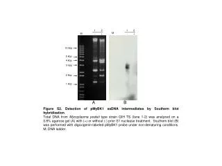
Blot hybridization of PCR products from several different follicular B cell lymphoma samples with MBI probe
Advantages of Southern blotting • Increases specificity of detection by incorporating two distinguishing features • Size • gel electrophoresis • Sequence • Complementary oligo- or polynucleotide probe • Increases sensitivity of fragment detection by using probe label that amplifies signal
Some Specific Purposes • To confirm that a PCR product includes a specific sequence • PCR product seen (or not seen) in gel contains expected sequence for a 14;18 translocation • To determine which fragment or fragments among the many resulting from a restriction enzyme digest contain a sequence of interest • To distinguish between a monoclonal and polyclonal population of B lymphocytes • To detect restriction fragment length polymorphisms (RFLPs) in genomic DNA
How much DNA should be run in a gel lane to allow visualization with probe? • Depends upon • the relative abundance of the target sequence to which hybridization must take place • the sensitivity of the visualization system • Current minimum = ~60fg of a 500-1000 bp band length • = to 60 fg of PCR product containing the sequence of interest (if using a polynucleotide probe) • can you see that with EtBr staining? • = to 120 ng of total human DNA to pick out a band of a single copy gene from a restriction digest • Radioactively labeled probe provides the greatest sensitivity, but for many applications, the sensitivity of non-radioactvely labeled probe is sufficient
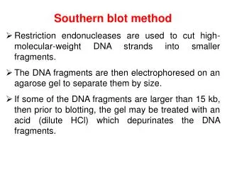
Prepare the DNA in the gel for blotting • Make DNA fragments >20 kb shorter so they blot out of the gel easily (but still in place). • nick by depurination • brief exposure to .25 – 0.5 N HCl • makes sugar-phosphate backbone open to cleavage by OH-. • Denature the DNA • soak gel in alkaline solution • makes DNA single-stranded so can hybridize with probe after blotting • neutralize following denaturation if doing a neutral transfer (more later)
Blot DNA to membraneMechanics of transfer • Capillary (no special equipment required!) • upward • downward • Electrophoretic • especially good for small fragments resolved by PAGE • Vacuum • more efficient and quantitative than capillary • must apply vacuum evenly and not too strongly
Transfer solutions - 3 main choices • Neutral, high ionic strength • Neutral, low ionic strength • Alkaline, low ionic strength
Transfer solution hints • Follow the membrane manufacturer’s recommendations • Alkaline blotting to charged nylon can increased background with chemiluminescent visualization. • Some nylon membranes deteriorate with lengthy exposure to alkaline conditions • Alkaline blotting doesn’t work with nitrocellulose • DNA won’t stick above pH 9 • Alkaline conditions degrade nitrocellulose • High ionic strength buffer works for all three membrane types, but alkaline to charged nylon is most efficient if background won’t be a problem!
How long does capillary transfer take?It depends on the • Size of DNA - the longer, the longer • % of agarose in gel - the higher, the longer • Thickness of the gel - the thicker, the longer • Direction of transfer - upward takes longer • accumulating pressure compresses gel and retards diffusion • Transfer buffer - • upward alkaline transfer takes ~2 hours • upward neutral transfer takes 12-24 hours
Membrane types • Charged nylon • Durable • Nylon modified with amine groups • Uncharged nylon • Durable • Nitrocellulose • Fragile • Used primarily for protein transfers
DNA fixation methods • Baking at 80oC • DNA non-covalently, hydrophobically bonded to any membrane • Alkaline blotting • DNA covalently bonded to charged nylon membrane • UV cross-linking • DNA covalently bonded to any nylon membrane
Blocking agents • SDS • Non-fat dry milk • BSA (bovine serum albumin) • PVP • Ficoll • Proprietary commercial preparations Every manufacturer of membranes makes specific blocking recommendations. It’s best to follow those first and modify as necessary.