Download
1 / 33
330 likes | 465 Views
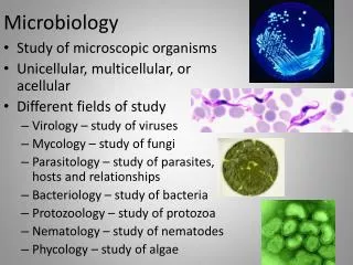
Microbiology. Chapter 16 Adaptive Immunity. Overview of Adaptive Immunity. Adaptive immunity is the body ’ s ability to recognize and defend itself against distinct invaders and their products Recognize specific sequences , not chemical patterns “Only” found in vertebrates

E N D
Microbiology Chapter 16 Adaptive Immunity
Overview of Adaptive Immunity • Adaptive immunity is the body’s ability to recognize and defend itself against distinct invaders and their products • Recognize specific sequences, not chemical patterns • “Only” found in vertebrates • Repeated contact with the same antigen results in more refined and faster response due to memory • The body constantly displays parts of proteins it is expressing (or finding) to show the adaptive immune system what type of proteins the host is holding • Some are “self” or “allowed to exist” and are tolerated • Remainders are foreign (“nonself”) and their sources and production eliminated
Five attributes of Adaptive Immunity • Specificity – each cell recognizes only a unique biochemical structure/sequence • Inducibility – needs some time to get fully activated • Need to amplify cells and create “improved” cells • Clonality – cells that recognize a pathogen are amplified • Recognizing a specific pathogen with a random sequence ~ hitting the jackpot or “getting lucky” • Those rare lucky cells need to be expanded because they are actually useful – used for refinement, memory, or specialized function to clear pathogens • Memory – cells that recognize a pathogen are stored, and back-up’s made for the next re-infection • Allows a rapid response upon reinfection • Unresponsiveness to self (or tolerance) • Cells that recognize self would be activated WAY too much to just be lucky; these are eliminated or “frozen”
Overview of Adaptive Immunity • Innate immune system “managed” by lymphocytes • Each manager is sequence-specific, so only a few are activated with each infection • These managers can activate innate immune cells to clear the infection • Two main types of adaptive lymphocytes • B lymphocytes (B cells) – mature in the bone marrow • Antigen-specific phagocytes • T lymphocytes (T cells) – mature in the thymus • Many managerial functions possible • Activate effector cells/ change how “cleanup” occurs • Activate B cells • Kill compromised tissue cells • Muffle/silence the “cleanup”
Two types of adaptive immune responses • Cell-mediatedimmune responses • Foreign particles localized and exactly defined • Intracellular infections – infections limited to specific cells • a few protozoa and bacteria • Virus-infected cells • nonself cells (tissue transplants; fetal cells; cancerous cells) • Eliminating the cell eliminates the pathogen • “surgical” cleansing – killing of cells; engulfing of cells and cell debris • Heavy use of macrophages and cytotoxic cells • inflammation and damage by friendly fire is often minimal • Humoralimmune responses • Foreign particles are diffuse or moving • Extracellular pathogens – infections can spread through tissues • Allergens, viruses between cells, released toxins • Heavy use of secreted molecules to tag pathogens (antibodies) or if necessary destroy everything in extracellular space (toxic enzymes) • Heavy use of granulocytes • “Carpetbombing” cleansing; inflammation and friendly fire common
Lymphocytes in motion https://www.youtube.com/watch?v=ntk8XsxVDi0 https://www.youtube.com/watch?v=UeW-lDmnl9M • Lymphocytes move like macrophages and amoeba • Note: • the amoeboid motion of the T cells • how the T cells “suction” onto target cells • how “excited” they get when they attach to a “matched” cell
How do lymphocytes learn about infections? • Lymphocytes are residents of the lymphatic system • Lymphocytes, being managers, sit at their desks (lymph nodes) and wait for the “action” to come to them • Most lymphocytes are resting in lymph nodes and do not move through the bloodstream • They leave their desks when they are “told” about infections, then enter the blood stream • Circulating lymphocytes are alerted to trouble and are trying to find the danger (to manage it)
About the Lymphatic System #1 • Tissues and Organs of the Lymphatic System • Purpose: cleanse interstitial fluid and return it to the blood • Lymph – interstitial fluid entering lymphatic vessels • Liquid with similar composition to blood plasma • Arises from fluid leaked from blood vessels into surrounding tissues • Contains: • salts and lipoproteins • cellular waste and debris • any pathogens and their debris from infections • Any metastatic cancers cells • dendritic cells from tissues with antigens looking for “matching” T cells • Lymphatic vessels • One-way system that drains interstitial fluid from the body tissues and returns it to the circulatory system • Run somewhat parallel to veins
Figure 16.2 The lymphatic system schematic 7200 L fluid/ day through 20 L fluid/ day into tissues 18 L fluid/ day out of tissues Note chains of lymph nodes leading to the subclabvian vein 2 L fluid/ day from tissues
About the Lymphatic System #2 • Tissues and Organs of the Lymphatic System • Lymphoid organs • Primary lymphoid organs – generate leukocytes • Red bone marrow – ALL cells, including early T cells • Thymus – T cell maturation and education • Secondary lymphoid organs – arranged in chains; filter fluids • “first” filters • Spleen – filters blood for pathogens • Tonsils – sense mouth, ear, eyes, sinus, nose fluid • Mucosa-associated lymphoid tissue (MALT) – sense gut and bronchioles • “downstream” filters • Lymph nodes – filter lymph from tissues and upstream lymphoid organs
Structure of secondary lymphoid organs • The spleen, tonsils, MALT, and lymph nodes are quite similar anatomically, and all have these structures: • Capsule – external covering; protrudes into structure via trabeculae • Nodules – units of lymph node segregated by trabeculae • Sinuses – channels for fluid flow through nodules • Cortex – outside layer • Paracortex – medial layer • Medulla – inside layer • Efferent lymphatic vessel – fluid exit from structure
Function of secondary lymphoid organs • Fluid flows into the structure • Mucus from oral/nasal cavity into tonsils • Mucus from gut/bronchioles into MALT • blood into spleen • Lymph from afferent lymphatic vessels into lymph nodes • Fluid flows first into the cortex of nodules, where they first interact with B cells. • Fluid next migrates to the paracortex to interact with T cells. • Fluid and any “matching‘” cells exit the lymph node at the medulla to head to the next node and eventually the general circulation.
Foreign particles: antigens vs. epitopes • Antigens • Properties of antigens • Molecules that elicit immune responses • Can initiate allergic reactions (allergens) • Most pathogens have many antigens for the immune system to detect • Food and dust can also contain antigenic (and allergenic) particles • Peanut, shellfish, gluten, pollen, mold • Epitopes • Portions of antigens that directly bind antibodies or T-cell receptors • What the immune system actually “sees” • Multiple epitopes occur on each antigen
Figure 16.3a Antigens, molecules that provoke a specific immune response. • Large foreign particles make the best antigens, especially if they have repeating units of epitopes • e.g., LPS in bacteria, capsomeres or spike proteins in viruses • loose or small antigens generally do not initiate strong immune responses (too few epitopes – poor resolution for immune system) • e.g., exotoxins, extracellular enzymes
Types of Antigens • Exogenous antigens – discovered outside of cells • includes toxins and other components of microbial cell walls, membranes, flagella, and pili • Immune system cells have to ingest them to learn about them • Endogenous antigens - discovered inside a body's cells • viral proteins, intracellular pathogens, mutated self-proteins (oncogenes) • Infected cells display epitopes for these antigens to immune system • Foreign antigens – by far the most common • Self antigens(autoantigens) – seen even when host is healthy – supposed to be ignored
T cells vs. B cells – SUMMARY SLIDE • Two groups of cells recognize specific sequences of amino acids or sugars • T Lymphocytes (T cells) - managers • They recognize only EPITOPES bound to MHC • When activated, they manage epitope-matched infections • T cells will NOT process any antigens unless they are infected • B Lymphocytes (B Cells) - workers • They recognize EPITOPES and phagocytose ANTIGENS in their natural form • They process these SPECIFIC antigens and present these SPECIFIC epitopes to T cells • B cells recognizing specific antigens make secreted forms of their receptors to bind antigen anywhere in the body • These secreted antigen-binding proteins are called antibodies
Pathogen Identification by B cells vs. T cells • Focus on what is holding the epitope, and how the cell behaves after the epitope is found
Elements of Adaptive Immunity • T Lymphocytes (T Cells) • Lifecycle: • Born in the red bone marrow • Mature in the thymus • Reside in secondary lymphoid tissues • Circulate between the lymph and blood • Have T cell receptors (TCRs) on their plasma membrane • Each TCR has a randomized receptor gene sequence • Each matured T cell will react to only a specific epitope, and only if bound to MHC • Most TCR’s never find their matched epitope • Other TCRs only recognize one epitope
How T cells detect epitopes • In all cells, endogenous antigens are broken down intracellularly • Antigen presenting cells (APCs) swallow exogenous antigens (pathogens) and break them down into epitopes within phagolysosomes • Dendritic cells – for initial detection in lymph nodes • Macrophages – for discovery in tissues after detection • B cells – pathogen specific – in lymph nodes and tissues • Some specialized endothelial cells – to hone to tissues • Protein epitopes are loaded onto “display” molecules called Major Histocompatibility Complexes (MHC) • Class I MHC displays endogenous epitopes found in all cells • Displays to active cytotoxic T cells • Class II MHC displays exogenous epitopes discovered by antigen-presenting cells (APCs) • Displays to helper, follicular, and regulatory T cells
Types of T lymphocytes • All develop from a single T-cell that “got lucky”, recognized a presented epitope, and subsequently expanded clonally • Memory T cells are generated and saved for re-infections • Four functional types of T cells based on (surface glycoproteins and) characteristic functions • Cytotoxic T lymphocyte • Directly kills compromised cells • Helper T lymphocyte • Directs effector immune response (macrophages, granulocytes, tissue cells) • Cell-mediated vs. humoral response to infection controlled here • Follicular T lymphocyte • Activates B cells at germinal centers of secondary lymphoid organs • Regulatory T lymphocyte • Represses adaptive immune responses • male-specific fetal antigens, prostate, brain antigens, ocular antigens, chronic mucosal resident microbia (gut, skin, oral, reproductive)
Magic of dendritic cells Dendrites used to increase contact area with T cells Also used to sample medium beyond tightly-bound cell borders (into lymph node sinuses, mouth, mucus, gut, epidermis, lungs)
Learning about an antigen • How B cells detect epitopes • Each B cell recognizes a distinct epitope because its B cell receptor (BCR) gene is randomized. • The BCR binds its specific epitope in lymph nodes or in tissues, almost always associated with its antigen • NO presentation molecules needed • B cells phagocytose the antigen and break it down into epitopes within phagolysosomes • The B cell loads all protein epitopes from the antigen onto MHC Class II, especially the epitopes it did not recognize • B cells are epitope-specific • Antigens have many epitopes • B cells cannot recognize any other epitopes, but can display ALL epitopes to T cells
B Lymphocytes (B Cells) and Antibodies • Found in clusters near T cells • primarily in the cortex of spleen, lymph nodes, tonsils, and MALT • Very few B cells circulate in the blood • Usually recently-activated B cells • Only a “lucky” B cell will recognize a foreign antigen • The B cell will expand into a population of thousands to millions at lymph node when it finds a matching T cell • Germinal center forms from expanding B cells and expanding matched T cells (swollen lymph node) • In some expanded B cells, The BCR is further randomized to try to make a tighter-binding BCR – affinity maturation • A soluble form of the “lucky” receptor is secreted to find, bind, and label antigen anywhere in the body – antibody • Many expanded B cells are terminally differentiated into short-lived plasma cells, that become antibody factories • Some are “back-ups” to be saved for future re-infections - memory B cells
Elements of Adaptive Immunity • Functions of Antibodies • Antigen-binding sites are complementary to epitopes • Antibodies function in several ways – “kisses of death” • Activation of complement and inflammation – through classical complement pathway • Neutralization – block up active sites of pathogenic enzymes or prevent critical contacts from happening • Opsonization – antibody-coated pathogens are tagged and are very easily discovered by the immune system • Agglutination – form a massive protein web that precipitates and stops pathogens from moving – easier to catch and swallow • Antibody-dependent cellular cytotoxicity (ADCC) – antibodies bound to a compromised host cell are ‘tagged’ for “assisted suicide” by cytotoxic immune cells
Elements of Adaptive Immunity • B Lymphocytes and Antibody Classes • “first guess” • All naïve and invariant B cells produce this antibody • IgM—first antibody produced; strongly activate complement; allow even weak pathogen contacts to trigger strong response (5x pathogen binding sites) • IgD—exact function is not known (membrane-bound form); mucosal immunity???? • “better guess” • Pathogen-experienced B cells produce these antibodies • IgA—associated with ALL body secretions • IgG—most common and longest-lasting antibody; transfers into mother’s milk and through placenta • IgE—involved in response to parasitic infections and allergies; granulocyte activators
Table 16.3 Characteristics of the Five Classes of Antibodies
Elements of Adaptive Immunity • Immune Response Cytokines • Soluble regulatory proteins that act as intercellular signals – coordinate immune system function during an infection and clearing of the infection and tissue repair afterward • Cytokines secreted by various leukocytes • They allow managers to control cells without having to touch them – “manager’s e-mails” • Cytokine network • Complex web of signals among cells of the immune system • “many e-mails”, effector cells decide course of action based on all cytokines present
Memory Cells and the Benefit of Immunological Memory • Primary immune response • Very few cells initially “match” the foreign antigens • Small amounts of antibodies produced initially • May take days to produce enough memory cells, plasma cells, and antibodies to eliminate the antigen from the body • Tends to be of low affinity, primarily IgM • Secondary (and subsequent) immune response • Far more memory cells already “matched” to re-exposed foreign antigens • Antibodies produced much faster • Tends to be high affinity antibody response • Tends to be mostly IgG
Figure 16.19 The production of primary and secondary antibody immune responses.
Types of Acquired Immunity • Specific immunity acquired during an individual’s life • Two types • Naturally acquired • Response against antigens encountered in daily life • Obtained through NATURAL sources • natural exposure to pathogen, placenta, mother’s milk • Artificially acquired • Response to antigens introduced via a vaccine • Distinguished as either active or passive • Active – the body generates its own response • Passive – the protection comes from the outside