Download
1 / 1
10 likes | 112 Views
Explore the analogy between human cochlea and a laser cavity, investigating the active amplification that facilitates hearing as well as solutions for sensorineural hearing loss. The study utilizes a state space formulation to test theories on the generation of spontaneous otoacoustic emissions (SOAEs) and examines the implications of perturbations in cochlear feedback gain. Gain insights into how variations in micromechanical parameters can lead to instability in cochlear models and compare the response of a nonlinear cochlea with that of a typical laser. By modeling both healthy and damaged cochleae, the aim is to enhance understanding of hearing mechanisms and develop interventions for the hearing impaired.

E N D
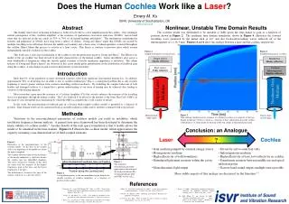
Does the Human Cochlea Work like a Laser?Emery M. KuISVR, University of Southampton, UKek@isvr.soton.ac.uk Abstract The sharply tuned sense of hearing in humans is believed to be due to active amplification in the cochlea. One seemingly natural consequence of this ‘cochlear amplifier’ is the existence of spontaneous otoacoustic emissions (SOAEs), narrow-band tones that are detected in the ear canals of 33% to 70% of all normal-hearing individuals.1 The mechanisms underlying the features and generation of SOAEs have long been a subject of debate. Zweig and Shera2 argue that SOAEs are created by multiple travelling-wave reflections between the middle ear boundary and a dense array of inhomogeneities scattered throughout the cochlea; Shera3 likens this process to activity in a laser cavity. This theory is contrary to previous ideas which assume independently unstable oscillators in the cochlea. This work uses a state space formulation of the cochlea to test the predictions made by Zweig and Shera2. The Elliott et al. model of the cat cochlea4 has been revised to describe characteristics of the human cochlea. Linear instabilities arise across a wide bandwidth of frequencies when the smooth spatial variation of basilar membrane impedance is disturbed. The salient features of Zweig and Shera’s theory2 are observed in this active model given perturbations in the distribution of feedback gain along the cochlea. A step change in gain is used to demonstrate system instability. Nonlinear, Unstable Time Domain Results The cochlear model was determined to be unstable at 2kHz given the step-change in gain as a function of position shown in Figure 2. The nonlinear time domain simulation, shown in Figure 4, illustrates the forward travelling wave generated by the initial click at t=0, and also backward-travelling waves reflected off of the inhomogeneity at x = 16.3 mm. Figures 4 and 6 show the analogy between a laser and the cochlea, respectively. Figure 4 Simplified diagram of a typical laser. Figure 6 Simplified diagram of a cochlea.. Output mirror, Mostly reflective Middle Ear, Mostly reflective Back mirror, Fully reflective Cochlear Inhomogeneity, Slightly reflective Introduction More than 8% of the population of many developed countries suffer from significant sensorineural hearing loss. In addition, approximately 90% of all hearing loss in adults is due to cochlear malfunction.5 This is a widespread problem that is only recently beginning to receive greater attention from scientists modelling cochlear mechanics. By modelling the complex behaviour of both healthy and damaged cochleae, it is hoped that a greater understanding of our sense of hearing may be achieved, thus leading to solutions for the hearing impaired. Most researchers agree upon the existence of a Cochlear Amplifier (CA) that actively enhances the response of the travelling wave as it propagates through the human cochlea. The CA is believed to be driven by the motility of the Outer Hair Cells (OHCs) in the organ of corti, though the exact mechanism by which the OHCs accomplish this is still a matter of debate. In this study, the micromechanical feedback gain in a discrete fluid-coupled cochlear model6 is perturbed as a function of position. Time-domain results are also generated from an unstable nonlinear cochlea and its attributes compared with a typical laser. Nonlinear Gain Medium Pumped Laser Medium Methods Figure 5 Time domain simulation of the response of a nonlinear cochlea to an impulse at the base. Basilar membrane velocity is shown as a function of time and position along the cochlea. A black line at 16.3 mm marks the location of the step change in micromechanical gain. Monochromatic lightoutput Narrow-band sound output Variations in the micromechanical parameters of cochlear models can result in instability, which invalidates frequency-domain analysis. A general state space framework has been developed to determine the linear stability of cochlear models.4 Another benefit of the state space formulation is that it readily allows the model to be simulated in the time-domain. Figures 1-3 illustrate the cochlear model, which approximates the organ by assuming a one-dimensional set of fluid-coupled elements. Conclusion: an Analogue ? Laser Cochlea Figure 1 Illustration of the macromechanics of the cochlear model. At the base of the cochlea (left) is the impedance of the middle ear, where the input is supplied. The cochlear elements represent the mechanics of the basilar membrane, a shelf that divides the cochlea into two fluid-filled chambers. The active and passive elements of this shelf are tuned such that response of the highest frequencies peak at the base, and the lowest frequencies at the apex. The helicotrema is located at the apex of the cochlea, and serves as a pressure release. • Gain medium pumped by external energy source • Homogeneous medium • High reflectivity at both boundaries • Stimulated light must resonate within the cavity • Monochromatic light output • Driven by active outer hair cells • Inhomogeneous medium • High reflectivity at base, low reflectivity in cochlea • Sound must resonate between middle ear and apical reflection point • Narrow-band sound output; multiple tones possible Figure 3 This nonlinear, micromechanical model3 approximates the motion of the basilar membrane (M1), tectorial membrane (M2), and OHC activity (γZ4). Figure 2 A step inhomogeneity in the micromechanical gain disturbs the smooth variation of cochlear impedance as a function of position in the cochlea. More subtle aspects of this analogy are discussed in the literature.6,7 References 1. Talmadge, C.L., Long, G.R., Murphy, W.J., Tubis, A. (1993). “New off-line method for detecting spontaneous otoacoustic emissions in human subjects,” Hear. Res., 71. 2. Zweig, G., and Shera, C.A. (1995). “The origin of periodicity in the spectrum of evoked otoacoustic emissions,” J. Acoust. Soc. Am. 98. 3. Shera, C.A., and Zweig, G. (1990). “Reflection of retrograde waves within the cochlea and at the stapes,” J. Acoust. Soc. Am. 89(3. 4. Elliott, S.J., Ku, E.M., and Lineton, B. (2007). “A state space model for cochlear mechanics,” J. Acoust. Soc. Am. 122(5), 2759-2771. 5 Jesteadt, W. (Editor) (1997). Modeling Sensorineural Hearing Loss. Hillsdale, NJ: Erlbaum. 6 S.T. Neely, D.O. Kim, (1986). A model for active elements in cochlear biomechanics. J. Acoust. Soc. Am. 79(5) 6. Shera, C.A. (2003). “Mammalian spontaneous otoacoustic emissions are amplitude stabilized cochlear standing waves,” J. Acoust. Soc. Am 114(1). 7. Ku, E.M., Elliott, S.J., and Lineton, B. (2008) “Statistics of instabilities in a state space model of the human cochlea,” submitted to J. Acoust. Soc. Am. for publication.


