Download
1 / 35
360 likes | 510 Views
Proteins: Evolution, and Analysis Lecture 7 9/15/2009. Chapter 4. (1). G A V L I M P F W. S T N Q Y C. Chapter 4. (2). K R H D E. (3). The Fischer Convention. Absolute configuration about an asymmetric carbon. related to glyceraldehyde (+) = D -Glyceraldehyde

E N D
Chapter 4 (1) G A V L I M P F W
S T N Q Y C Chapter 4 (2)
K R H D E (3)
The Fischer Convention Absolute configuration about an asymmetric carbon related to glyceraldehyde (+) = D-Glyceraldehyde (-) = L-Glyceraldehyde
Cahn - Ingold - Prelog system Can give absolute configuration nomenclature to multiple chiral centers. Priority Atoms of higher atomic number bonded to a chiral center are ranked above those of lower atomic number with lowest priority away from youR highest to lowest = clockwise, S highest to lowest = counterclockwise SH>OH>NH2>COOH>CHO>CH2OH>C6H5>CH3>H
Newman Projection • A projection formula representing the spatial arrangement of bonds on two adjacent atoms in a molecular entity. • The structure appears as viewed along the bond between these two atoms, and the bonds from them to other groups are drawn as projections in the plane of the paper. • The bonds from the atom nearer to the observer are drawn so as to meet at the centre of a circle representing that atom. • Those from the further atom are drawn as if projecting from behind the circle.
The major advantage of the CIP or RS system is that the chiralities of compounds with multiple asymmetric centers can be unambiguously described
Overview of Protein Sequencing (1) Purify Protein (2) Determine number of PP End group analysis (Dansyl chloride rxn) (3) Fragment PP into smaller peptides Enzymes (Trypsin, Chymotrypsin, etc.) Chemical (CNBr) (4) Determine the sequence Edman Degradation with PITC (5) Assemble a sequence (6) Elucidate S-S bonds Amino acid composition
Long peptides have to be broken to shorter ones to be sequenced
Q9. You must cleave the following peptide into smaller fragments. Which of the proteases listed in the table would be likely to yield the most fragments? The fewest? NMTQGRCKPVNTFVHEPLVDVQNVCFKE
Reconstructing the protein’s sequence Specific chemical cleavage reagents Cleave the large protein using i.e trypsin, separate fragments and sequence all of them. (We do not know the order of the fragments!!) Cleave with a different reagent i.e. Cyanogen Bromide, separate the fragments and sequence all of them. Align the fragments with overlapping sequence to get the overall sequence.
How to assemble a protein sequence 1. Write a blank line for each amino acid in the sequence starting with the N-terminus. 2. Follow logically each clue and fill in the blanks. 3. Identify overlapping fragments and place in sequence blanks accordingly. 4. Make sure logically all your amino acids fit into the logical design of the experiment. 5. Double check your work.
1 2 3 4 5 6 7 8 9 10 11 12 13 14 15 H3N+-_-_-_-_-_-_-_-_-_-_-_-_-_-_-_-COO- A-T F - M - A - T A- K - F - M Q - M -A - K D - I - K - Q - M G - M - D - I - K Y - R - G - M Y - R Cyanogen Bromide (CNBr) Cleaves after Met i.e M - X D - I - K - Q - M A - T A - K - F - M Y - R - G - M Trypsin cleaves after K or R (positively charged amino acids) Q - M - A - K G - M - D - I - K F - M - A - T Y - R
Q11. Separate cleavage reactions of a polypeptide by CNBr and chymotrypsin yield fragments with the following amino acid sequences. What is the the sequence of the intact polypeptide? • CNBr treatment Chymotrypsin 1. Arg-Ala-Tyr-Gly-Asn 1. Met-Arg-Ala-Tyr 2. Leu-Phe-Met 2. Asp-Met-Leu-Phe 3. Asp-Met 3. Gly-Asn Q13. Treatment of a polypeptide with 2-mercaptoethanol yields two PP: 1. Ala-Val-Cys-Arg-Thr-Gly-Cys-Lys-Asn-Phe-Leu 2. Tyr-Lys-Cys-Phe-Arg-His-Thr-Lys-Cys-Ser Treatment of the intact PP with trypsin yields fragments with the following aa compositions: 3. (Ala, Arg, Cys2, Ser, Val) 4. (Arg, Cys2, Gly, Lys, Thr, Phe) 5. (Asn, Leu, Phe) 6. (His, Lys, Thr) 7. (Lys, Tyr)
Sequencing by Mass Spectrometry Electrospray Ionization Mass Spectrometry ESI-MS spectrum of horse heart apomyoglobin Q. Two successive peaks in the mass spectrum have measure m/z ratios of 1414.0 and 1542.3. What is the original apomyoglobin molecule? p1= (M+z)/z p2= (M+z-1)/z-1 M= 16,975D (16,951 D in table 5-1)
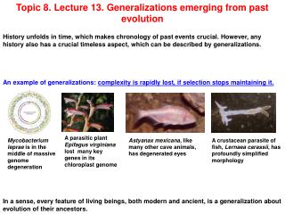
Protein Evolution Species variation in homologous proteins The primary structures of a given protein from related species closely resemble one another. If one assumes, according to evolutionary theory, that related species have evolved from a common ancestor, it follows that each of their proteins must have likewise evolved from the corresponding ancestor. A protein that is well adapted to its function, that is, one that is not subject to significant physiological improvement, nevertheless continues to evolve. Neutral drift: changes not effecting function
Homologous proteins (evolutionarily related proteins) Compare protein sequences: Conserved residues, i.e invariant residues reflect chemical necessities. Conserved substitutions, substitutions with similar chemical properties (Asp for Glu), (Lys for Arg), (Ile for Val) Variable regions, no requirement for chemical reactionsetc.
Man,chimp0 Rhesus monkey 1 0 Average differences Horse 12 11 0 Donkey 11 10 1 0 10.0 cow,sheep 10 9 3 2 0 dog 11 10 6 5 3 0 gray whale 10 9 5 4 2 3 0 5.1 rabbit 9 8 6 5 4 5 2 0 kangaroo 10 11 7 8 6 7 6 6 0 Chicken 13 12 11 10 9 10 9 8 12 0 penguin 13 12 12 11 10 10 9 8 10 2 0 9.9 Duck 11 10 10 9 8 8 7 6 10 3 3 0 14.3 Rattlesnake 14 15 22 21 20 21 19 18 21 19 20 17 0 12.6 turtle 15 14 11 10 9 9 8 9 11 8 8 7 22 0 Bullfrog 18 17 14 13 11 12 11 11 13 11 12 11 24 10 0 Tuna fish 21 21 19 18 17 18 17 17 18 17 18 17 26 18 15 0 18.5 worm fly 27 26 22 22 22 21 22 21 24 23 24 22 29 24 22 24 0 silk moth 31 30 29 28 27 25 27 26 28 28 27 27 31 28 29 32 14 0 25.9 Wheat 43 43 46 45 45 44 44 44 47 46 46 46 46 46 48 49 45 45 0 Bread mold 48 47 46 46 46 46 46 46 49 47 48 46 47 49 49 48 41 47 54 0 47.0 Yeast 45 45 46 45 45 45 45 45 46 46 45 46 47 49 47 47 45 47 47 41 0 Candida k. 51 51 51 50 50 49 50 50 51 51 50 51 51 53 51 48 47 47 50 42 27 0 Man,chimp monkey Horse Donkey cow,sheep dog gray whale rabbit kangaroo Chicken, penguin Duck Rattlesnake turtle Bullfrog Tuna fish worm fly silkworm Wheat Bread mold Yeast Candida Amino acid difference matrix for 26 species of cytochrome c
Phylogenetic tree • Indicates the ancestral relationships among the organisms that produced the protein. • Each branch point indicates a common ancestor. • Relative evolutionary distances between neighboring branch points are expressed as the number of amino acid differences per 100 residues of the protein. • PAM units • or • Percentage of Accepted Mutations
PAM values differ for different proteins. Although DNA mutates at a assumed constant rate. Some proteins cannot accept mutations because the mutations kill the function of the protein and thus are not viable.
Mutation rates appear constant in time Although insects have shorter generation times that mammals and many more numbers of replication, number of mutations appear to be independent of the number of generations but dependent upon time Cytochrome c amino acid differences between mammals, insects and plants note the similar distances
Evolution through gene duplication • Many proteins within an organism have sequence similarities with other proteins. • These are called gene or protein families. • The relatedness among members of a family can vary greatly. • These families arise by gene duplication. • Once duplicated, individual genes can mutate into separate genes. • Duplicated genes may vary in their chemical properties due to mutations. • These duplicate genes evolve with different properties. • Example the globin family.
Genealogy of the globin family • Hemoglobin: • is an oxygen transport protein • it must bind and release oxygen as the cells require oxygen • Myoglobin: • is an oxygen storage protein • it binds oxygen tightly and releases it when oxygen concentrations are very low a2b2 a2g2 a2d2 z2e2
The globin family history 1. Primordial globin gene acted as an Oxygen-storage protein. 2. Duplication occurred 1.1 billion years ago. lower oxygen-binding affinity, monomeric protein. 3. Developed a tetrameric structure two a and two b chains increased oxygen transport capabilities. (a2b2). Mammals have fetal hemoglobin with a variant b chain i.e. g (a2g2). 5. Human embryos contain another hemoglobin (z2e2). 6. Primates also have a d chain with no known unique function. (a2d2).
Modular Construction of some proteins Modules (sequence motifs): ~ 40 -100 residues
Lecture 8 (9/17/2009) Chapter 6 - Proteins: 3-D structure 6-1. Secondary Structure