Download
1 / 1
10 likes | 137 Views
B cell trafficking in Bovine Leukemia Virus infected sheep.

E N D
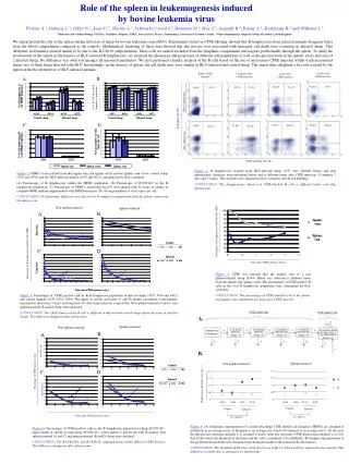
B cell trafficking in Bovine Leukemia Virus infected sheep Debacq Christophe 1*, Florins Arnaud 1*, Asquith Becca 2, Gillet Nicolas 1*, Sanchez-Alcaraz Maria Teresa 1, Boxus Mathieu 1, Urbain Patrice 1, Schwartz-Cornil Isabelle 3, Bonneau Michel 3, Jean Geneviève 4, Kerkhofs Pierre 5, Hay Jack 6, Théwis André 4, Kettmann Richard 1*, Willems Luc 1* 1. Molecular and Cellular biology, FUSAG, Gembloux, Belgium; 2. Department of Immunology, Imperial College, London, UK; 3. U892 INRA, Jouy-en-Josas, France; 4. Zootechnie, FUSAG, Gembloux, Belgium; 5. Department of Virology, Veterinary and Agrochemical Research Centre, Uccle, Belgium; 6. Department of Laboratory Medicine and Pathobiology University of Toronto, Toronto, Canada * are members of the FNRS. Lymphocyte homeostasis within the peripheral blood results from a critical balance between proliferation and cell renewal. Disruption of this subtle equilibrium consecutively to an increase in cellular proliferation and/or a reduction of cell death can potentially lead to the onset of leukemia. Based on intravenous bromodeoxyuridine injection, we previously demonstrated that the proliferation rate was increased approximately two-fold in BLV infected sheep (2 % are produced by proliferation per day versus 1.1 % in controls), whereas the death rate of these proliferating cells remained unchanged (Debacq et al., 2002 Proc. Natl. Acad. Sci. U.S.A. 99: 10048-10053). The imbalance created between proliferation and death rates would potentially lead to a cell population growth of 7 % in the first day. Although this phenomenon might occur in some acute leukemic phases, the cell numbers usually remain rather constant in chronically infected animals. Therefore, enhanced proliferation, which is thought to occur in lymphoid tissues, should be counterbalanced either by differential recirculation (i.e. homeostasis) or by increased cell death in other organs (or both). We tested these hypotheses by labeling the peripheral blood lymphocytes with carboxyfluorescein diacetate succinimidyl ester (CFSE) and tracing their migration through the lymph nodes. No significant difference in cell kinetics was observed in the efferent lymphatics of BLV-infected and control sheep, indicating that their migration efficiency was unaltered. In the peripheral blood, however, the proportion of the CFSE-positive cells decreased faster in BLV-positive animals. Considering that CFSE-labeled proteins are equally distributed upon cell division, mathematical calculations of the turnover parameters showed that the death rates were increased while proliferation was unchanged. In other words, BLV infection of sheep is characterized by increased proliferation in lymphoid organs (i.e. more frequently proliferating cells) concomitantly with enhanced death in the periphery (i.e. faster disappearance of the total blood cell population). Interestingly, splenectomy abrogated the difference in kinetics observed between infected and control sheep, demonstrating major role exerted by the spleen in modulating B-cell dynamics. 1 A. 2 A. 3. 2 B. 1 B. Figure 1: B cell trafficking through the lymph node A. Intestinal or prescapular efferent lymphatic from BLV-infected (n° 4535) and control (n° 4533) sheep were surgically cannulated allowing chronic sampling of lymph. Animals were next injected intravenously with 25 mg of carboxyfluorescein diacetate succinimidyl ester (CFSE) dissolved in 4 ml of dimethylsulfoxide (DMSO) containing 10 U/µl of heparin. At regular intervals of time (6, 22 and 94 hours), lymph was collected, B cells were labeled with 1H4 monoclonal antibody and stained with a phycoerythrin (PE)-conjugated antibody. Then, ten thousand cells were analyzed by two-color flow cytometry (x axis = CFSE; y axis = B-lymphocytes). The percentages of B cells labeled with CFSE are indicated in the upper right quadrants. B. Lymph samples from three BLV-infected (n° 4535, 4536 and 1095) and three control sheep (n° 4533, 4534 and 2152) were collected at different days post-CFSE injection and the mean percentages of CFSE-positive cells within the total B-lymphocyte population were determined. Figure 3: Kinetic analyses of B cells subsets At regular intervals of time, peripheral blood mononuclear cells (PBMCs) were isolated from two BLV-infected (n° 4535 and 4536) and two control sheep (n° 4333 and 4534). Cells were next labeled with a B-lymphocyte specific antibody and with a peridinin-chlorophyll-protein (PerCp)-conjugated secondary antibody. Then, the PBMCs were incubated with monoclonal antibodies directed against L-selectin, CD21, CD5 or CD11b, and with a PE-conjugated antibody. Finally, thrice-labeled cells (CFSE/PE/PerCP) were analyzed by flow cytometry. Percentages of B cell subsets labeled with CFSE are represented at different time intervals (in days). Figure 2: B cell kinetics in the blood A. One BLV-infected (n° 4535) and one control (n° 4533) sheep were injected intravenously with CFSE and an aliquot of blood was collected by jugular venipuncture 15 minutes, 46 hours and 5 weeks after the injection. After red blood cell were lysis, B cells were labeled with 1H4 monoclonal antibody, stained with a PE-conjugated antibody and analyzed by flow cytometry (x axis = CFSE; y axis = B-lymphocytes). Ten thousand cells (lymphocytes, monocytes and granulocytes) were acquired. The percentages of B cells labeled with CFSE are indicated in the upper right quadrants. B. Short-term (4 days) and long-term kinetics of the CFSE-positive B cell percentages in the circulating blood of two BLV-infected (n° 4535 and 4536) and two control sheep (n° 4533 and 4534). 4 A. 5 A. 5 B. 4 B. Figure 5: Mathematical modelling of the CFSE data A. Schematic representation of a model describing CFSE labeled cell dynamics. PBMCs are assumed to proliferate at an average rate p, to disappear at an average rate d and to be replaced at an average rate L. On division, the fluorescence intensity (initially J) is assumed to halve. After five divisions, CFSE fluorescence intensity is so low that it falls below the threshold of detection and the cell is considered to be unlabeled. B. Graphic representations of the minimal proliferation and death rates estimated from fitting the model to the measured B cells kinetics. In non-splenectomized sheep, the p values are statistically similar between the infected (n° 4535 and 4536) and the control sheep (n° 4533 and 4534) (p>0.05 according to the two tailed Mann-Whitney test). However, the d values are statistically different between the two groups of animals (p<0.05 according to the two tailed Mann-Whitney test). In splenectomized sheep, the p and d values are statistically similar between the BLV-infected (n° 4535 and 3002) and the uninfected splenectomized sheep (n° 4534 and 3004) (p>0.05 according to the two tailed Mann-Whitney test). Figure 4: Effect of splenectomy on the B cell dynamics A. Percentages of CFSE-positive B cells in the circulating blood B lymphocyte population of two splenectomized BLV-infected (n° 4535 and 3002) and two splenectomized control sheep (n° 4534 and 3004). B. Percentages of CFSE-positive cells in B lymphocytes expressing or not CD11b in two splenectomized BLV-infected (n° 4535 and 3002) and two splenectomized control sheep (n° 4534 and 3004).