Download
1 / 83
840 likes | 1.01k Views
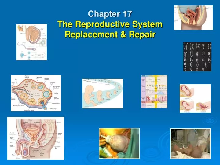
Chapter 17 The Reproductive System Replacement & Repair. Introduction Purpose of Reproduction. Mitosis or Asexual reproduction: Tissue Replacement Regeneration of Tissues Cells & tissues get damaged or simply wear out.

E N D
IntroductionPurpose of Reproduction Mitosis or Asexual reproduction: Tissue Replacement • Regeneration of Tissues • Cells & tissues get damaged or simply wear out. • The process by which cells make exact copies of themselves, & is essential to maintain a healthy body. Meiosis: • To reproduce human life
Mitosisvs Meiosis Mitosis: • Cells make identical copies of themselves • Human cells are called Eukaryotic cells • Contain bundles of chromatin (chromosomes) #46 • Bacteria are single-cell Prokaryotic cells & divide via Binary Fission. Prophase Metaphase Anaphase Telophase & Cytokinesis
Meiosis Gametes: • Sex cells; female eggs & male sperm for human or animal reproduction. • Formed in Gonads: Ovaries & Testes • Total number of chromosomes needed is 46, 23 from mother & 23 from father. • Daughter Cells: produced at end of meiosis have half as many chromosomes (23) as original. • Chromosomes are matched based on size, shape, & genes they carry & numbered 1-23.
Meiosis • 23rd pair: is sex chromosome determines sex of baby. • XX is female, XYis male; thus the father determines sex of the baby because the mother can onlycontribute “X” chromosomes.
Sexual Reproduction • Fertilization: both male & female gametes unite & combine their genetic material; thus fertilization. • Zygote (fertilized egg), has 46 chromosomes; reproduces millions of times via mitosis & develops within female to change from embryo to fetus.
The Early Stages of the Human Life Cycle • Day 1: Fertilization in Fallopian tube • Week 1-3: Zygote in Fallopian tube • Weeks 3-9: Embryo implantation in uterus • Week 10: Fetus placenta with umbilicus formed • Month 9: Infant birth
The Human Reproduction SystemFemale Anatomy • Reproductive organs called genitalia Divided into: • Primary genitalia: gonads (Ovaries) that produce gametes. • Secondary genitalia: other structures that aid in reproductive process; fallopian tubes, uterus, vagina, & external genitalia called vulva.
The Ovary • Covered by fibrous capsule called tunica albuginea made of cuboidal epithelium. • Cortex: contains eggs (gamates) • Medulla: contains blood vessels, nerves, & lymphatic tissue surrounded by loose connective tissue.
The Uterine Tubes • Calledoviducts or fallopian tubes, are 4 inch passageways for eggs to get to uterus. • Infundibulum: large funnel, surrounded by ciliated projections called fimbria. • Connected to superior portion of uterus • Made of smooth muscle, ciliated simple columnar epithelium tissue. • Covered by visceral peritoneum & suspended by mesentery known as mesosalpinx, Latin for tuba!!!
The Uterus • Found in pelvic cavity posterior and superior to urinary bladder & anterior to rectum. • Major portion called “body.” • Fundus: rounded superior portion between uterine tubes. • Isthmus: narrow inferior portion • Cervix: “valve-like” portion of uterus that protrudes into vagina. • Cervical Canal: communicates with vagina
Uterine Layers • Perimetrium: outermost layer • Myometrium: smooth muscle • Endometrium: highly vascular inner lining is mucosal layer of columnar epithelium & secretory cells with two divisions; 1. Basal: responsible for regenerating uterine lining each month. 2. Functional: sheds about every 28 days during menses.
Pathology Connection: Endometriosis Etiology: endometrial tissue escapes uterus & implants in abdominal cavity. Builds up & decays each month with hormonal changes. S/S: • Chronic pelvic pain • dysmenorrhea: pain during menstruation • dyspareunia: painful sexual intercourse • Dysuria: painful urination
Endometriosis con’t DX: • history & physical exam • ultrasound • laparoscopic surgery Rx: • NSAIDs • surgical removal of tumors • Hormones: progesterone, GnRH agonists, androgens (testosterone). • Hysterectomy
The Vagina • 10 cm (4 inches) long running from uterus to outside of body. Purpose: • To receive penis during intercourse • Allow for passage of menstrual fluid out of uterus • Also called birth canal, to allow movement of baby out of uterus during childbirth. • Opening may be covered a perforated membrane, the hymen.
The External Genitalia (Vulva) Labia majora: • rounded fat deposits that meet & protect rest of external genitalia. • meet anteriorly to form “mons pubis” Pudendal cleft: • Open area between the 2 labia majora
The External Genitalia (Vulva) con’t Perineum: • Area between vagina & rectum Labia minora: (prepuce) • Meet anteriorly to form the prepuce • Clitoris contains erectile tissue 2cm in diameter
Mammary Glands • Milk production glands housed in breasts • In young children virtually identical in boys & girls • At puberty estrogen & progesterone stimulate breast development in girls.
Lactation • In adult females, breast consists of 15-20 glandular lobes & “adipose” tissue. • Each lobe divided into smaller lobules which house milk secreting sacs called alveoli when woman is “lactating.” • Milk travels through series of ducts & sinuses, eventually reaching areola or nipple. • Milk production controlled by hormone “prolactin” (AP)
The Menstrual Cycle • Organized around approximately 28 days, involves ovaries & uterus. • Ovarian cycle involves monthly maturation & release of eggs from ovary. • Uterine cycle consists of monthly buildup, decaying, & shedding of uterine lining.
The Menstrual Cycle con’t • Cycles begin in teen years during puberty, & end during her 40s or 50s in menopause. • Goal is to release egg for fertilization, prepare uterus to receive fertilized egg, & nourish fertilized egg should pregnancy result. • If pregnancy doesn’t result, uterine lining will shed & cycle will begin again!
The 28-dayMenstrual Cycle • Begins with first day of menses • Menstruation refers to actual shedding of endometrium, “period” itself. • Menses usually lasts4–5 days, but can be longer or shorter in different women, can vary month to month.
The 1st of 4 Stages of Menses Follicle Stage: • 10 days long • FSH released (AP) • Follicle & ovum mature • Estrogen released (ovary) • Uterine lining prepared
The 2nd of 4 Stages of Menses Ovulation Stage: • FSH stops • LH starts: triggers ovulation (AP) Day 14 • Follicle ruptures • Ovum released
The 3rd of 4 Stages of Menses Corpus Luteum Stage: • Progesterone released (ovary & endometrium) • If egg fertilized, progesterone cont • 14 days long
The 4th of 4 Stages of Menses Menstruation Stage: • If no egg fertilized, 3-6 days of diminished production of progesterone. • Uterine lining broken down
Oogenesis, Follicle Development, & Ovulation Oogenesis: • Process of egg production • Begins with birth of Oogonia, or egg stem cells, in ovary. • Oogonia undergoes mitosis, producing millions of “primary oocytes”. • Women will have all eggs they will ever have 5 months before they are born.
Primary Oocytes • Born via mitosis: still have 46 chromosomes • Must undergo meiosisto become gametes, with only 23 chromosomes, allowing them to combine with another gamete (sperm) & ending with total of 46 chromosomes. • Primary oocytes stay in kind of suspended animation until puberty, when they finish developing.
Follicle Development • Primary oocytes eventually surrounded by helper cells, called granulosa cells; at this point they are called primordial follicles, & stay “dormant” until puberty. • Hormonal signals during puberty cause some primordial follicles to enlarge & increase number of granulosa cells. • Enlarged cells called “primary follicles”
Ovulation • @ puberty, one primary follicle will become “secondary” follicle. • Secondary follicle: will not complete development unless it is ovulated & fertilized. • Just before ovulation: secondary follicle fills with fluid & moves toward surface of ovary, where it becomes a visible lump.
Ovulation con’t • Fimbria of uterine tubes brush surface of ovary, causing follicle to rupture. • When follicle ruptures: egg (actually an oocyte) is released into peritoneal cavity & fimbria pull it toward funnel, drawing it into uterine tube. (Wow!!!)
Ovulation con’t • Ova travels down Fallopian tube • If sperm present in uterine tube & all conditions are right, sperm will penetrate egg, fertilizing it & triggering rest of egg development.
If ova is fertilized… • Now called “zygote” with 46 chromosomes • Zygote enters uterus & implants into proliferated “endometrium,” stopping woman from experiencing menses. • Ruptured follicle left behind in ovary will become “corpus luteum” and secrete hormones to help maintain thickened endometrium to nourish embryo.
If ova is NOT fertilized… • zygote will not implant in uterus • Within few days, uterine lining will begin to degenerate & woman will have her period, with egg swept out in menstrual flow. • Corpus luteum will become corpus albicans & eventually disappear.
Hormonal Control • Hormones from hypothalamus, pituitary, & ovary control female cycle. • Generally controlled bynegative feedback loop. • Released as part of hierarchy, with hypothalamus releasing hormone that controls pituitary, which then releases hormone that controls another organ.
The 4 hormones that control menstrual cycle • Ovary: Estrogen & Progesterone • Pituitary: Luteinizing hormone (LH) & Follicle Stimulation Hormone (FSH). • Estrogen & progesterone levels: increase at puberty. • Gonadotropin releasing hormone (GnRH) from hypothalamus causes increase in secretion of LH & FSH from pituitary.
Hormonal Controlcon’t During Follicular stage: FSH initiates development of primary follicles each month, while LH triggers ovulation. • Estrogen levels continue to rise as more & more is secreted by developing follicle, stimulating proliferation of uterine lining. • Estrogen exerts positive influence on hypothalamus, increasing secretion ofGnRH, thus increasing LH & FSH. • + feedback loop continues, raising hormone levels until ovulation occurs.
Hormonal Controlcon’t • Once ovulation occurs, feedback loop reverses itself. • Corpus luteum begins to secrete progesterone, as well as a little estrogen. • Estrogen under influence of progesterone, exerts neg. feedback on hypothalamus & pituitary, decreasing GnRH, LH, & FSH secretion.
Hormonal Control con’t • Progesterone also exerts negative feedback on hypothalamus & pituitary. • Thus, LH, FSH, & estrogen levels drop while progesterone levels rise. • Hormonal changesprevent another egg from maturing.
Hormonal Control con’t • For about 10 days after ovulation, progesterone levels remain high as corpus luteumcontinues to secrete hormone. • Progesterone's effect on uterus is to maintain buildup of endometrium & to decreaseuterine contractions. • If no pregnancy results, corpus luteumdegenerates & progesterone will no longer be produced.
Hormonal Control con’t • Decreasing progesterone levels cause degeneration of endometrium, followed by menstruation. • Decreased progesterone levels release hypothalamus & pituitary from its inhibitory effects, & FSH & LH levels begin to rise & cycle begins again.
If pregnancy occurs… • Implanted fertilized egg secretes hormone called human chorionic gonadotropin (HCG). • HCG stimulates corpus luteum to keep secreting progesterone & little estrogen to maintain uterine lining. • @ about three month’s gestation, placenta begins to secrete its own progesterone & estrogen, thus becoming endocrine organ!!!
If pregnancy occurs… con’t… • Prolactin: produced by anterior Pituitary stimulates milk production by breasts. • Oxytocin: produced by posterior Pituitary stimulates uterine contractions & milk production.
Pathology Connection: Menstrual Disorders Premenstrual Syndrome: Etiology: central-nervous-system neurotransmitter interactions with sex hormones affected??? S/S: depression, anger, irritability, anxiety, confusion, withdrawal, breast tenderness, bloating, swelling extremities, headache. D/X: must start 5 days prior to menses, cease 4 days into menses, & not recur until following cycle; X 3 cycles.
Premenstrual Syndrome con’t Rx: • Mild/moderate: good nutrition, regular exercise, complex carbohydrates, calcium supplements. • NSAID orhormonal suppression • Severe: psychiatric medications
Polycystic Ovarian Syndrome Etiology: Ovaries produce too much testosterone & estrogen, but too little progesterone, effects 5-10 % women. S/S: • Failure to ovulate • Excess facial hair • Male pattern baldness • Irregular menstrual cycles • Formation of multiple cysts on ovaries • Hormonal abnormalities • Infertility • Obesity • Insulin resistance (Type II DM)
Polycystic Ovarian Syndrome con’t D/X: • Serum analysis; looking for elevated levels of LH,testosterone, prolactin, insulin. • Ultrasound: looking for large ovaries Rx: consists mainly of symptom relief • Weight loss • Birth control pills: can regulate menstrual cycle • Drugs: can treat hyperlipidemia& DM • Infertility drugs &/or surgical repair of damaged ovaries.
Breast Cancer • Etiology: most common malignancy of American women and leading cause of death between ages 40-55; familial history • S/S: bloody, brown, or serous nipple discharge, noticeable lump, changes in breast tissue • DX: mammogram, Imaging, biopsy • TX: lumpectomy, mastectomy, chemotherapy, radiation, etc