Diaphragm
Diaphragm. Ajith Sominanda Department of Anatomy Faculty of Medicine. Objectives. Gross structure, function & dysfunction 1 . Describe the component parts of the diaphragm and state its functions 2. Describe the nerve supply and blood supply of the diaphragm

Diaphragm
E N D
Presentation Transcript
Diaphragm Ajith Sominanda Department of Anatomy Faculty of Medicine
Objectives Gross structure, function & dysfunction 1. Describe the component parts of the diaphragm and state its functions 2. Describe the nerve supply and blood supply of the diaphragm 3. State and identify structures passing through the diaphragm including the vertebral levels 4. Use the knowledge of Anatomy in examining the respiratory system 5. State and describe the common clinical problems of the thoracic cavity 6. Clinical correlations of diaphragm related to respiration Development 1. Development of the respiratory system and associated developmental abnormalities 2. Describe the development of the diaphragm including its congenital abnormalities
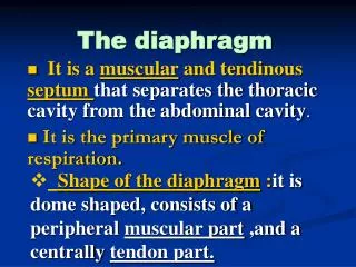
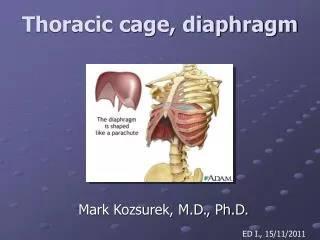
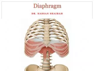
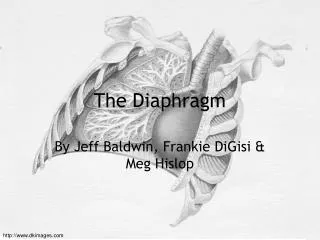
What is the Diaphragm • Fibromuscular / musculo-tendinous structure that separates the thoracic cavity from the abdominal cavity. • Main function is inspiration • Developed from inner (transversus) layer of body wall muscles
Other diaphragms in human body • Floor of the mouth – Oral diaphragm • Thoracic diaphragm- The diaphragm • Floor of the pelvis – Pelvic diaphragm
Components Domes or Cupola of diaphragm Tendinous part (central tendon)x Muscular part Base of the diaphragm
Attachments • Xiphoid process anteriorly (T8-T9) • Costal margin of the thoracic wall • Ends of ribs XI and XII; • Ligaments & crura that span across structures of the posterior abdominal wall • Vertebrae of the lumbar region. Median arcuate ligament Medial arcuate ligaments Lateral arcuate ligaments
Structures pass through T8 T10 T12
Blood supply • Costal margin: lower five intercostal and subcostal arteries • Muscle fibers arising from crura: Left & right inferior phrenic arteries from abdominal aorta • Other contributions: pericardiacophrenic & musculophrenic from internal thoracic artery superior phrenic from thoracic aorta
Nerve supply • Motor: • Phrenic nerve (C3,4,5) • Sensory (proprioceptive) • Costal margin by intercostal nerves • Central area by phrenic nerves
Actions • Inspiration / Breath in • Abdominal straining
Clinical correlation: Level and the area around of the diaphragm
Clinical correlation: Examination of level of diaphragm Diaphragmatic hernia • Abnormal opening in the diaphragm specially during birth i.e. congenital • Abdominal contents herniate into thorax causing respiratory problems