Thoracic cage, diaphragm
700 likes | 3k Views
Thoracic cage, diaphragm. Mark Kozsurek, M.D., Ph.D. ED I., 15/11/2011. Questions to be answered How are the bones of the thorax connected together? → the joints of the thoracic cage Which muscles act upon these joints? → the muscles of the thoracic cage How does the thoracic cage move?

Thoracic cage, diaphragm
E N D
Presentation Transcript
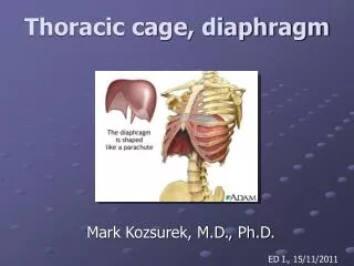
Thoracic cage, diaphragm Mark Kozsurek, M.D., Ph.D. ED I., 15/11/2011
Questions to be answered • How are the bones of the thorax connected together? → the joints of the thoracic cage • Which muscles act upon these joints? → the muscles of the thoracic cage • How does the thoracic cage move? → the mechanism of breathing
How are the ribs connected to the vertebrae? How do the ribs attach to the sternum?
How are the ribs connected to the vertebrae? COSTOVERTEBRAL JOINTS: • 1. COSTOTRANSVERSE JOINT • - costal fovea of the transverse process • articular facet of the the costal tubercle 2. JOINT OF HEAD OF RIB - costal facet of the vertebra - articular facet of the head of the rib
Articular surfaces of vertebrae • „TYPICAL VERTEBRAE” • TII-IX present a superior and an inferior costal demifacet. • „ATYPICAL VERTEBRAE” • - TI bears a complete superior costal facet for the head of the first rib and an inferior costal demifacet for the head of the second rib. • TX only has a superior costal demifacet. • TXI-XII express complete costal facets for the ribs with the same number. They have no costal facets on their transverse processes. TYPICAL
Articular surfaces of the ribs Ribs II-X Rib I Ribs XI-XII Ribs I, XI-XII have continuous facets on their heads, the articular surfaces of the heads of ribs II-X are divided into two by a crest, ribs XI-XII have no tubercle and do not articulate with transverse processes.
Joint of the head of the rib Costotransverse joint
Radiate ligament of the head of the rib (not shown) Joint of the head of the rib Costotransverse joint
lateral costotransverse lig. superior costotransverse lig. radiate lig. of the head of the rib
Costotransverse ligament attaches the neck of the rib to thetransverse process. Lateral costotransverse ligament connects the tip of the transverse process to the roughened nonarticular part of the tubercle of the rib. Superior costotransverse ligament attaches the neck of the rib to the transverse process of the vertebra above. • Intra-articular ligament connects the crest of the head of the rib to the intervertebral disc. Radiate ligament of the head of the rib reinforces the capsule of the joint of the head of the rib.
How are the ribs connected to the sternum? Manubriosternal and xiphisternal joints: synchondroses, with age they become ossified. Sternocostal joints: Rib I: synchondrosis Ribs II-VII: synovial joints Intercostal joints: Ribs VI-X synovial joints
external intercostal m. internal intercostal membrane internal intercostal m. innermost intercostal m. transversus thoracis m. external intercostal membrane vertebra rib sternum
Posterior intercostal arteries are direct branches of the thoracic aorta, while the anterior intercostal arteries arise from the internal thoracic artery, a branch of the subclavian artery. Supreme intercostal arteries of the two upper intercostal spaces also come from the subclavian artery. • Posterior intercostal veins drain into the azygos vein (on the right) or into the hemiazygos/ accessory hemiazygos veins (on the left). Anterior intercostal veins open into the internal thoracic vein, which empties into the brachiocephalic vein. • Intercostal nerves exit from the spinal cord and leave the vertebral canal through the intervertebral foramina. • Intercostal vessels and nerves give off posterior, lateral and anterior branches to supply the external layers of the thoracic wall.
1. 4. 2. 3. 5. • Pectoralis major (and minor) • Serratus anterior • Abdominal muscles • Trapezius (Levator scapulae, Rhomboids, Serratus posterior) • Latissimus dorsi
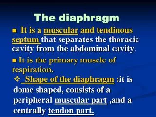
Central tendon with the opening for the inferior vena cava a b b According to the site of origin muscle fibres constitute the: a, sternal part b, costal part c, lumbar part IVC esophagus aorta c c Diaphragm superior aspect
sternal part costal part lumbar part sternocostal triangle (of LARREY) lumbocostal triangle (of BOCHDALEK)
medial part lateral part lateral, medial, median arcuate ligaments L1 L2 L3
Esophageal opening: esophagus, ant. et post. vagal trunk, T10
Aortic hiatus: aorta, thoracic duct (the greatest lymph vessel), L1
Sternocostal triangle of Larrey: internal thoracic artery and vein * * they are called superior epigastric a. et v. bellow the diaphragm
Between the medial and lateral parts (variable!!!): greater splanchnic nerve, azygos/hemiazygos veins, lesser splanchnic nerve and sympathetic trunk
Internal thoracic artery (ITA) is one of the first branches of the subclavian artery. ITA gives off the pericardiacophrenic artery first which descends together with the phrenic nerve between the pleura and the pericardium. Before reaching the trigone of Larrey, ITA gives off the musculophrenic artery contributing to the blood supply of the diaphragm. • Already from the abdominal aorta the inferior phrenic arteries ascend to the inferior surface of the diaphragm. • Phrenic nerve arises mainly from the C4 segment, this is why injuries of the neck around or above this level may cause sudden death due to the palsy of the diaphragm.
Respiratory movements Ribs rotate around an axis defined by the two costovertebral joints.
During inspiration both the anteroposterior and lateral diameters of the chest increase!
Quiet breathing Inspiration (active) Expiration (passive) Note: The position of the central tendon and the heart resting on it is not changing during quiet breathing!!! In midposition (green) the right dome of the diaphragm reaches the level of the 5th rib, while on the left it is in the 5th intercostal space.
90° External and internal intercostal muscles are perpendicular to each other!
external intercostal internal intercostal
Take a deep breath and relax! It’s over!