Download
1 / 23
230 likes | 275 Views
Explore the structure, regions, and functions of the spinal cord, a vital link between the brain and body. Learn about spinal nerves, reflex arcs, dermatomes, nerve plexuses, and reflex testing in a clinical setting. Understand how spinal cord injuries and abnormalities manifest in clinical applications.

E N D
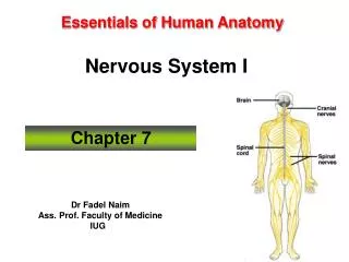
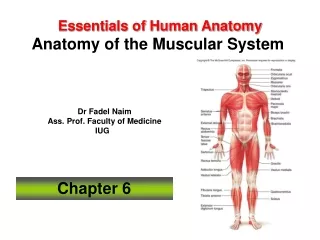
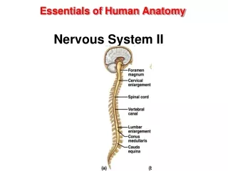
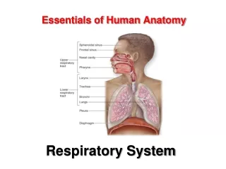
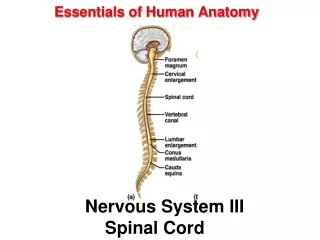
Essentials of Human Anatomy Nervous System III Spinal Cord
The Spinal Cord • Link between the brain and the body. • Exhibits some functional independencefrom the brain. • The spinal cord and spinal nerves serve two functions: • pathway for sensory and motor impulses • responsible for reflexes
Structure of the Spinal Cord • Typical adult spinal cord • ranges between 42 and 45 centimeters (cm) in length. • In cross section • roughly cylindrical • slightly flattened both posteriorly and anteriorly. • External surface has twolongitudinal depressions: • the posterior (dorsal) median sulcus • the anterior (ventral) median fissure
Regions of the Spinal Cord • The cervical region • continuous with the medulla oblongata • contains neurons whose axons form the cervical spinal nerves (8) • The thoracic region • attached to this region are the thoracic spinal nerves (12) • The lumbar region • contains the neurons for the lumbar spinal nerves (5) • The sacral region • contains the neurons for the sacral spinal nerves (5) • The coccygeal region • one pair of coccygeal spinal nerves arises from this region
Structure of the Spinal Cord • The spinal cord is shorter than the vertebral canal that houses it. • Conus medullaris: • tapered inferior end of the spinal cord • marks the official “end” of the spinal cord proper. • Cauda equina • Inferior to conus medularis • nerve roots (groups of axons) that project inferiorly from the spinal cord. • Filum terminale • Within the cauda equina • thin strand of pia mater • helps anchor the conus medullaris to the coccyx.
Spinal Meninges • Are continuous with the cranial Meninges. • Structures that encircle the spinal cord, listed from superficial to deep are: • vertebra • epidural space • Dura mater • subdural space • arachnoid • subarachnoid space • pia mater
Spinal Nerves • 31 pairs • connect the CNS to: • receptors • muscles, glands • Each spinal nerve is mixed: • thousands of motor and sensory axons. • Sensory axons originate from receptors • Motor axons originate from the spinal cord. • Anterior root and posterior root unite within the intervertebral foramen • become a spinal nerve. • Spinal nerve is associated with the vertebra of the same number.
Rami of Spinal Nerves • Posterior (or Dorsal) ramus • Innervates muscles and skin of the back • Anterior Ramus • Largest branch • Forms plexuses • Innervates anterior and lateral trunk, upper and lower limbs
Spinal Nerves • Dorsal root (posterior or sensory root) • axons of sensory neurons in the dorsal root ganglion • Ventral root (anterior or motor root) • axons of motor neurons whose cell bodies are in spinal cord • Dorsal root ganglion • cell bodies of sensory neurons whose axons conduct impulses inward from peripheral body parts • Spinal nerve • union of ventral root and dorsal root 11
Dermatomes • A specific segment of skin supplied by a single spinal nerve. • All spinal nerves • innervate a segment of skin and are associated with a dermatome. • except for C1 • Dermatome map: • sensory segments: skin of the body associated with a spinal nerve
Intercostal Nerves • Anterior rami of spinal nerves T1–T11. • Travel in the intercostal space sandwiched between two adjacent ribs
Nerve Plexuses • A network of interweavinganterior rami of spinal nerves. • nerve plexuses on both the right and left sides of the body. • Nerve plexuses then split into multiple “named” nerves that innervate various body structures. • Principal plexuses • cervical plexuses • brachial plexuses • lumbar plexuses • sacral plexuses.
Reflexes • Fast, stereotypical, inborn, protective actions • Occur at spinal cord or brainstem levels • May be either monosynaptic or polysynaptic • All require a. stimulus at receptor b. sensory information relay c. processing at CNS level d. activation of motor response e. response of peripheral effector
Reflex Arcs Reflexes – automatic, subconscious responses to stimuli within or outside the body 18
Monosynaptic Reflexes • The simplest of all reflexes. • No interneurons. • The patellar (knee-jerk) reflex is a monosynaptic reflex
Polysynaptic Reflexes • Have more complex neural pathways • exhibit a number of synapses • involve interneurons within the reflex arc. • Has more components • more prolonged delay between stimulus and response.
Reflex Testing in a Clinical Setting • Reflexes can be used to test specific muscle groups and specific spinal nerves or segments of the spinal cord. • Consistently abnormal reflex response may indicate damage to the nervous system or muscles. • A reflex response may be normal, hypoactive, or hyperactive.
Clinical Application Cerebral Injuries and Abnormalities • Concussion • brain jarred against cranium • loss of consciousness • temporary loss of memory • mental cloudiness • headache • recovery usually complete • Cerebral Palsy • motor impairment at birth • caused by blocked cerebral blood vessels during development • seizures • learning disabilities • Cerebrovascular Accident • stroke • sudden interruption in blood flow • brain tissues die 22