Download
1 / 70
730 likes | 955 Views
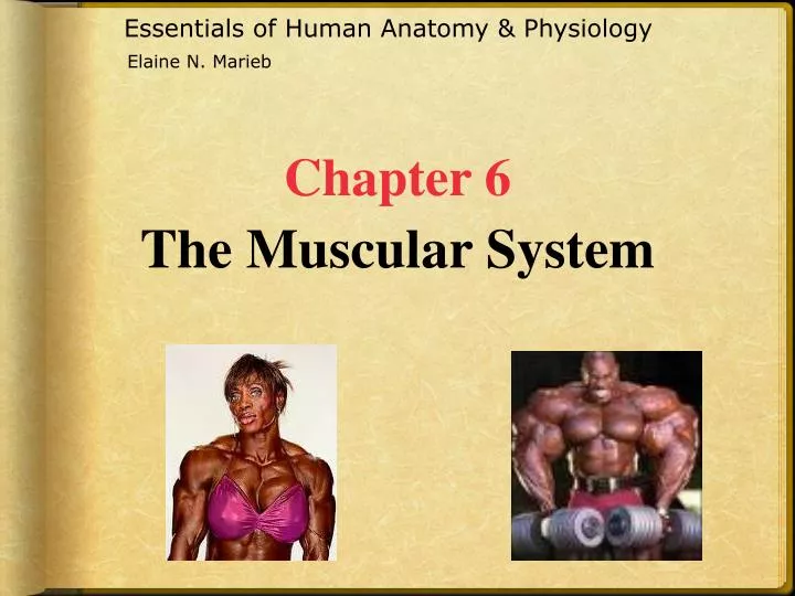
Essentials of Human Anatomy & Physiology. Elaine N. Marieb. Chapter 6 The Muscular System. The Muscular System. Muscles are responsible for all types of body movement – they contract or shorten and are the “machines” of the body Three basic muscle types are found in the body Skeletal muscle

E N D
Essentials of Human Anatomy & Physiology Elaine N. Marieb Chapter 6The Muscular System
The Muscular System • Muscles are responsible for all types of body movement – they contract or shorten and are the “machines” of the body • Three basic muscle types are found in the body • Skeletal muscle • Cardiac muscle • Smooth muscle
Characteristics of Muscles • Skeletal & smooth muscle cells are elongated (muscle cell = muscle fiber) • Contraction of muscles is due to the movement of microfilaments • All muscles share some terminology • Prefix myo refers to muscle • Prefix mys refers to muscle • Prefix sarco refers to flesh
Skeletal Muscle Characteristics • Most are attached by tendons to bones • Cells are multinucleated • Striated – have visible banding • Voluntary – subject to conscious control • Cells are surrounded and bundled by connective tissue = great force, but tires easily
Connective Tissue Wrappings ofSkeletal Muscle • Endomysium – around a single muscle fiber • Perimysium – around a fascicle (bundle) of fibers
Connective Tissue Wrappings ofSkeletal Muscle • Epimysium – covers the entire skeletal muscle • Fascia – on the outside of the epimysium
Skeletal Muscle Attachments • Epimysium blends into a connective tissue attachment • Tendon – cord-like structure • Aponeuroses – sheet-like structure • Sites of muscle attachment • Bones • Cartilages • Connective tissue coverings
Smooth Muscle Characteristics • Nostriations • Spindle-shaped cells • Single nucleus • Involuntary – no conscious control • Found mainly in the walls of hollow organs • Slow, sustained contractions (tireless)
Cardiac Muscle Characteristics • Has striations • Usually has a single nucleus • Joined to another muscle cell at an intercalated disc • Involuntary • Found only in the heart • Steady pace!
Muscle Functions • Produce movement • Maintain posture • Stabilize joints • Generate heat
Microscopic Anatomy of Skeletal Muscle • Cells are multinucleate • Nuclei are just beneath the specialized plasma membrane called Sarcolemma
Microscopic Anatomy of Skeletal Muscle • Myofibril • Bundles of myofilaments • Myofibrils are aligned to give distinct bands (striations) • I band = light band • A band = dark band
Microscopic Anatomy of Skeletal Muscle • Sarcomere • Contractile unit of a muscle fiber
Microscopic Anatomy of Skeletal Muscle • Organization of the sarcomere • Thick filaments = Myosin filaments • Composed of the protein myosin • Contain ATPase enzymes • Extend the entire length of the dark A band
Microscopic Anatomy of Skeletal Muscle • Myosin filaments • Myosin heads • Create cross bridges
Microscopic Anatomy of Skeletal Muscle • Organization of the sarcomere • Thin filaments = Actin filaments • Composed of the protein actin • Anchored to the Z disc
Microscopic Anatomy of SkeletalMuscle • Sarcoplasmic reticulum • Specialized smooth endoplasmic reticulum • Stores and releases calcium on demand when the muscle fiber is stimulated to contract
Skeletal Muscle Activity • Stimulation & Contraction of Single Skeletal Muscle Cells • Irritability – ability to receive and respond to a stimulus • Contractility – ability to shorten when an adequate stimulus is received
Nerve Stimulus to Muscles • Skeletal muscles must be stimulated by a nerve to contract (motor neruron) • Motor unit • One neuron • Muscle cells stimulated by that neuron
Nerve Stimulus to Muscles • Neuromuscular junctions – association site of nerve and muscle
Nerve Stimulus to Muscles • Synaptic cleft – gap between nerve and muscle • Nerve and muscle do not make contact • Area between nerve and muscle is filled with interstitial fluid
Transmission of Nerve Impulse to Muscle • Neurotransmitter – chemical released by nerve upon arrival of nerve impulse • The neurotransmitter for skeletal muscle is acetylcholine • Neurotransmitter attaches to receptors on the sarcolemma • Sarcolemma becomes permeable to sodium (Na+)
Transmission of Nerve Impulse to Muscle • Sodium rushing into the cell generates an action potential • Once started, muscle contraction cannot be stopped
The Sliding Filament Theory of Muscle Contraction • Activation by nerve causes myosin heads (crossbridges) to attach to binding sites on the thin filament • Myosin heads then bind to the next site of the thin filament
The Sliding Filament Theory of Muscle Contraction • This continued action causes a sliding of the myosin along the actin • The result is that the muscle is shortened (contracted)
Contraction of a Skeletal Muscle • Muscle fiber contraction is “all or none” • Within a skeletal muscle, not all fibers may be stimulated during the same interval • Different combinations of muscle fiber contractions may give differing responses • Graded responses – different degrees of skeletal muscle shortening, rapid stimulus = constant contraction or tetanus
Muscle Response to Strong Stimuli • Muscle force depends upon the number of fibers stimulated • More fibers contracting results in greater muscle tension • Muscles can continue to contract unless they run out of energy
Energy for Muscle Contraction • Initially, muscles used stored ATP for energy • Bonds of ATP are broken to release energy • Only 4-6 seconds worth of ATP is stored by muscles • After this initial time, other pathways must be utilized to produce ATP
Energy for Muscle Contraction • Direct phosphorylation • Muscle cells contain creatine phosphate (CP) • CP is a high-energy molecule • After ATP is depleted, ADP is left • CP transfers energy to ADP, to regenerate ATP • CP supplies are exhausted in about 20 seconds
Energy for Muscle Contraction • Anaerobic glycolysis • Reaction that breaks down glucose without oxygen • Glucose is broken down to pyruvic acid to produce some ATP • Pyruvic acid is converted to lactic acid
Energy for Muscle Contraction • Anaerobic glycolysis (continued) • This reaction is not as efficient, but is fast • Huge amounts of glucose are needed • Lactic acid produces muscle fatigue
Energy for Muscle Contraction • Aerobic Respiration • Series of metabolic pathways that occur in the mitochondria • Glucose is broken down to carbon dioxide and water, releasing energy • This is a slower reaction that requires continuous oxygen
Muscle Fatigue and Oxygen Debt • When a muscle is fatigued, it is unable to contract • The common reason for muscle fatigue is oxygen debt • Oxygen must be “repaid” to tissue to remove oxygen debt • Oxygen is required to get rid of accumulated lactic acid • Increasing acidity (from lactic acid) and lack of ATP causes the muscle to contract less
Types of Muscle Contractions • Isotonic contractions • Myofilaments are able to slide past each other during contractions • The muscle shortens • Isometric contractions • Tension in the muscles increases • The muscle is unable to shorten
Muscle Tone • Some fibers are contracted even in a relaxed muscle • Different fibers contract at different times to provide muscle tone • The process of stimulating various fibers is under involuntary control
Effects of Exercise on Muscles • Results of increased muscle use • Increase in muscle size • Increase in muscle strength • Increase in muscle efficiency • Muscle becomes more fatigue resistant
Five Golden Rules of Skeletal Muscle Activity With a few exceptions, all muscles cross at least one joint Typically, the bulk of the muscle lies proximal to the joint crossed All muscles have at least 2 attachments origin & insertion Muscles can only pull; they never push During contraction, the muscle insertion moves toward the origin
Muscles and Body Movements • Movement is attained due to a muscle moving an attached bone
Muscles and Body Movements • Muscles are attached to bone, or to other connective tissue structures, at no less than two points • Origin – attachment to the stationary bone • Insertion – attachment to the movable bone
Types of Body Movements • Flexion- Decreases the angle of the joint and brings 2 bones closer together (Bending the knee or elbow) • Extension – Opposite of flexion- increases the angle between 2 bones (Straightening the knee or elbow) • Hyperextension- Extension >180 degrees (Tipping your head back so your chin points toward the ceiling)
Types of Body Movements Rotation- Movement of a bone around its longitudinal axis; Common with ball and socket joints describes the movement with the atlas around the dens of the axis (Shaking your head “no”) Abduction – Moving a limb away from the midline of the body; Fanning movement of fingers, toes Adduction – Movement of a limb towards the body midline; Opposite of abduction Circumduction – Combination of flexion, extension, abduction and adduction. Proximal end of limb is stationary, distal end moves in a circle Limb as a whole outlines a cone See textbook page 195
Left: Abduction – moving the leg away from the midline Right: Circumduction: cone-shaped movement, proximal end doesn’t move, while distal end moves in a circle. Above : Adduction- moving toward the midline
Special Movements • Dorsiflexion Lifting the foot so that its superior surface approaches the shin • Plantar flexion Pointing the toes • Inversion To invert the foot- turn the sole medially • Eversion To evert the foot- turn the sole laterally
Special Movements • Supination- Forearm rotates laterally so that the palm faces anteriorly, and the radius and ulna are parallel (Carry “soup” --> “soup”-inating) • Pronation- Forearm rotates medially so the palm faces posteriorly (Face down) • Opposition- Action by which you move your thumb to touch the tips of the other fingers on the same hand (See textbook page 195)