Download

1 / 44
440 likes | 720 Views
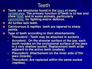
Identifying Structures, Teeth, and Names of Teeth Units 18.1 – 18.2 Dr. Hale. Medical Technologies Jr. Program. A. Odontology is the study of the anatomy, growth, and diseases of the teeth B. Teeth are accessory organs of the digestive tract that aid in the mastication, or chewing, of food

E N D
Identifying Structures, Teeth,and Names of TeethUnits 18.1 – 18.2Dr. Hale Medical Technologies Jr. Program
A. Odontology is the study of the anatomy, growth, and diseases of the teeth B. Teeth are accessory organs of the digestive tract that aid in the mastication, or chewing, of food C. Individuals have two dentitions or sets of teeth
1. Primary or deciduous dentition a. First set of teeth b. At birth, a newborn has about 44 teeth buds at various stages of development c. Teeth buds begin to erupt into the mouth at about 6 months of age Eruption of teeth - YouTube
d. When a child is about 2 years old, all the 20 primary or deciduous teeth have erupted e. Functions 1) Maintain proper spacing for the permanent teeth 2) Used for mastication and speech
2. Permanent or succedaneous dentition a. Second set of teeth b. When primary teeth are lost between the ages of 6 and 12 years, they are replaced by permanent or succedaneous teeth c. Permanent teeth begin to erupt at about 5 years of age
d. Continue erupting and replacing primary teeth until about 17 to 20 years of age when the third molars or wisdom teeth erupt e. Most of the 32 teeth in permanent dentition are in place by 12 years of age
3. Mixed dentition a. Occurs in a child 5 to 12 years old b. Both primary and permanent teeth erupted in the mouth
Sections or Divisions of a Tooth 1. Four main sections or divisions: crown, root, cervix, and apex 2. Crown a. Section of the tooth that is visible in the mouth b. Protected on the out- side by a tissue called enamel
3. Root a. Section of the tooth below the gingiva or gums b. Covered on the outside by a tissue called cementum c. Normally not visible in the mouth d. Helps anchor or hold tooth in the bony socket of the jaw e. Tooth may have a single root or multiple roots 1) Two roots: bifucated 2) Three roots: trifurcated
4. Cervix a. Also called the neck or cemento-enamel junction b. Narrow section where the crown joins with the root c. Area where enamel covering crown meets cementum covering root
5. Apex a. Tip of the root of the tooth b. Contains an opening called apical foramen through which nerves and blood vessels enter the tooth
Tissues of the Tooth 1. Each tooth is made of four main tissues: enamel, cementum, dentin, and pulp 2. Enamel a. Hardest tissue in the body b. Covers the outside of the crown c. Made up of mainly calcium and phosphorus d. Forms a protective layer for the tooth e. Once tooth is fully developed, enamel cannot grow or repair itself
3. Cementum a. Hard, bone-like tissue that covers outside of the root b. Provides a thin layer of protection c. Helps hold the tooth in place d. Cementum is formed throughout the life of the tooth
4. Dentin a. Tissue that makes up the main bulk of the tooth b. Bonelike substance but it is softer than enamel but harder than cementum that provides its outer coverings c. It has no nerves, it carries sensations of pain and temperature to the pulp
d. Dentin is a living tissue 1) Capable of limited repair and continued growth 2) Internal surface of dentin forms the wall of the pulp chamber
5. Pulp a. Soft tissue located in the innermost area of the tooth b. Made of blood vessels and nerves held in place by connective tissue c. Section of pulp located in crown is called pulp chamber
d. Section located in toot is called the pulp canal e. Pulp chamber and pulp canal create a space in the center of the tooth known as the pulp cavity f. Provides sensation and nourishment for the tooth g. Assists in the production of dentin
Periodontium 1. Consists of those structures that surround and support teeth 2. Includes alveolar process, periodontal ligament, and gingiva 3. Alveolar process or ridge
a. Bone tissue of the maxilla and mandible that surrounds the roots of the teeth b. Contains a series of sockets or alveoli, one for each tooth in the mouth c. Although the tooth sits in the alveolus and is supported by it, the tooth does not touch the bone
4. Periodontal ligament a. Dense fibers of connective tissue b. Attaches to the cementum and to the alveolus c. Supports or suspends the tooth in the socket
d. Acts as a shock absorber and prevents the tooth from resting on or rubbing against the bone during the chewing process e. Also contains nerves and blood vessels 1) Provide nourishment 2) Aid in the production of cementum 3) Produce sensation when pressure is applied to tooth
5. Gingiva or gums a. Made of epithelial tissue covered with mucous membrane b. Cover the alveolar bone and surround the teeth c. Free gingiva 1) Gingiva that surrounds the cervix of a tooth 2) Fills interproximal spaces 3) It is not attached to the tooth
4. Molars a. Teeth in back of mouth b. Largest and strongest teeth c. Used to chew and grind food
Primary or Deciduous Teeth 1. First set of teeth 2. Also called “baby” teeth a. This is poor term because it implies they are not important b. Serve important function of maintaining correct spacing for the permanent teeth 3. 20 teeth in all: 10 maxillary and 10 mandibular
4. 10 maxillary deciduous teeth a. Central incisors: 2 b. Lateral incisors: 2 c. Cuspids: 2 d. 1st molars: 2 e. 2nd molars: 2
5. 10 mandibular deciduous teeth a. Central incisors: 2 b. Lateral incisors: 2 c. Cuspids: 2 d. 1st molars: 2 e. 2nd molars: 2 6. There are no bicuspids in primary or deciduous dentition
7. Each of the teeth is labeled as maxillary or mandibular a. Maxillary: teeth are in the sockets or alveoli of the maxilla, or upper jaw bone b. Mandibular: teeth are in alveoli of the mandible, or lower jaw bone
8. Each tooth is also identified as right or left a. Positions are shown as though you are looking into another person’s open mouth b. Your left hand is by the patient’s right teeth c. Your right hand is by the patient’s left teeth
9. Each tooth has a specific name a. Label as right or left b. label as maxillary or mandibular c. Examples: 1) Maxillary right central incisor 2) Maxillary left central incisor 3) Mandibular right central incisor 4) Mandibular left central incisor d. Same system is used to name lateral incisors, cuspids, and molars
Permanent or Succedaneous Teeth 1. Second or permanent set of teeth 2. 32 teeth total: 16 maxillary and 16 mandibular 3. 16 maxillary or upper teeth a. Central incisors: 2 b. Lateral incisors: 2 c. Cuspids: 2
d. 1st bicuspids: 2 e. 2nd bicuspids: 2 f. 1st molars: 2 g. 2nd molars: 2 h. 3rd molars (wisdom teeth): 2
4. 16 Mandibular or lower teeth a. Central incisors: 2 b. Lateral incisors: c. Cuspids: 2 d.1st bicuspids: 2
e. 2nd bicuspids: 2 f. 1st molars: 2 g. 2nd molars: 2 h. 3rd molars (wisdom teeth): 2
5. Each tooth has a specific name a. Label each tooth as right or left b. Label each tooth as maxillary or mandibular c. Label each tooth with proper name
d. Examples 1) Maxillary right central incisor 2) Maxillary left central incisor 3) Mandibular right central incisor 4) Mandibular left central incisor e. Same system is used to name lateral incisors, cuspids, bicuspids, and molars