Tdp1-/- Mice Exhibit Aberrant Transcript Production and Altered Processing of Tyrosyl DNA Ends
50 likes | 167 Views
This study investigates the genomic alterations in Tdp1 knockout (Tdp1-/-) mice, focusing on the effects of exon deletions and the resulting aberrant transcripts. PCR analysis reveals distinct genomic DNA products for knockout and wild-type mice, while Southern blotting confirms the deletion of specific exons. Furthermore, real-time PCR demonstrates decreased transcript levels for specific exons in Tdp1-/- tissues. The processing capabilities of Tdp1-/- fibroblasts are assessed, highlighting deficiencies in 3'-tyrosyl processing, further elucidating Tdp1's role in genomic stability.

Tdp1-/- Mice Exhibit Aberrant Transcript Production and Altered Processing of Tyrosyl DNA Ends
E N D
Presentation Transcript
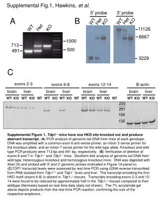
Supplemental Fig.1, Hawkins, et al. 5′ probe 3′ probe Het KO A WT Het KO WT B KO Het WT 11126 6667 1000 713 500 491 3229 C exons 2-3 exons 6-8 exons 12-14 B-actin brain liver brain liver brain liver brain liver WT KO WT KO NT WT KO WT KO NT WT KO WT KO NT WT KO WT KO 220 201 154 [AJH1]Need better supplemental figure legend title [AJH2]Need to fill this in. 134 Supplemental Figure 1.Tdp1-/-mice have one HKD site knocked out and produce aberrant transcript. (A) PCR analysis of genomic tail DNA from mice of each genotype. DNA was amplified with a common exon 8 anti-sense primer, an intron 5 sense primer for the knockout allele, and an intron 7 sense primer for the wild-type allele. Knockout and wild-type PCR products were 713 bp and 491 bp, respectively. (B) Verification of deletion of exons 6 and 7 in Tdp1+/- and Tdp1-/- mice. Southern blot analysis of genomic tail DNA from wild-type, heterozygous knockout and homozygous knockout mice. DNA was digested with XbaI (X) and probed with 5′ and 3′ genomic probes (indicated in Figure 1A panel iv). (C)TDP1 transcript levels were assessed by real time PCR using cDNA reverse transcribed from RNA isolated from Tdp1+/+ and Tdp1-/- brain and liver. The transcript encoding the first HKD motif (exons 6-8) is absent in Tdp1-/- tissues. Transcripts encoding exons 2-3 and 12-14 were found to be reduced by approximately half in the Tdp1-/- tissues compared to their wildtype littermates based on real-time data (data not shown). The 7% acrylamide gel above depicts products from the real-time PCR reaction, confirming the size of the respective amplicons.
Supplemental Fig.2, Hawkins, et al. Single-strand 18 nt 3'-phosphotyrosine *pTCCGTTGAAGCCTGCTTTp-Tyr 3' 5' A Tdp1+/+ Tdp1+/- Tdp1-/- none B pTyr p C Tdp1+/+ Tdp1+/- Tdp1-/- none pTyr p D Mouse Liver Homogenate Supplemental Figure 2. Tdp1‑/‑ mice are deficient in 3' tyrosyl processing. A radiolabeled (*) 3'-pTyr oligomeric substrate was treated with tissue homogenates from a Tdp1+/+, Tdp1+/‑, or Tdp1‑/‑mouse for 1 h, subjected to denaturing gel electrophoresis, and phosphorimaged. (A) Diagram of 3'-pTyr 18-nucleotide oligomeric substrate. The substrate was treated with four-fold serial dilutions of brain (B), or five-fold serial dilutions of liver (C) homogenate from a Tdp1+/+, Tdp1+/‑, or Tdp1‑/‑mouse. The percent conversion from the tyrosyl substrate to its phosphate productwas calculated by densitometry and shown for liver (D) homogenates.
Supplemental Fig.3, Hawkins, et al. A B repair products LV-IRES-DsRed LV-FLAGhTDP1 LVTHM-DsRed uninfected -Red 100 no extract 75 anti-FLAG Western pTyr OH -/- +/+ +/- MEF extracts Supplemental Figure 3. Tdp1+/‑ fibroblast cell line can also effectively process 3' tyrosyl DNA ends and Tdp1‑/‑ fibroblasts were genetically complemented via lentivirus infection. The substrate indicated in Figure 3A was treated with (A) 40, 20, or 10 μg of whole-cell extracts from Tdp1+/+, Tdp1+/‑, or Tdp1‑/‑MEF cell lines for 1 h. 15 and 30 μL of each lentiviral-infected Tdp1‑/‑MEFs lysate, harvested and identically prepared, were analyzed via Western blotting with (B) anti-FLAG antibody.
Supplemental Fig. 4, Hawkins et al. A no gap, nicked one base gap 25 17 * 25 17 * p p 42 43 = PG B 0 mM Mg, 5 mM EDTA + 2 mM Mg 10 mM Mg repair products untreated untreated untreated KO WT WT KO WT KO 35 17OH 17PG time C repair products 0 mM Mg, 5 mM EDTA 10 mM Mg 2 mM Mg untreated untreated untreated WT WT KO KO KO WT 17OH 17PG time
Supplemental Figure 4. Tdp1‑/‑ fibroblasts are not deficient in processing PG on SSBs. TDP1 can also process 3′‑PG termini, and under some conditions appears largely responsible for their removal [1]. MEF whole‑cell extracts were incubated with radiolabeled substrates that mimic SSBs bearing phosphoglycolate end modifications to assess the removal of this 3′ blocking moiety in the ‑/‑ MEFs . (A) Diagram of 3'‑PG SSB substrates; asterisk represents the 5′ radioactive tag on the 3'‑PG 17‑mer modification. Both the 42-base (B) and 43-base (C) substrates were treated with 20 μg of whole-cell extract from Tdp1+/+ and Tdp1‑/‑ cell lines for 5, 20, and 60 min in reaction buffer with 0, 2, and 10 mM Mg++. These conditions are either prohibitive (0 Mg++)or permissive (2, 10 mM Mg++) for the activity of Ape1 [2] or PNKP. In (B), rightmost lane contains radiolabeled marker of 35 bases. Generation of fully repaired 42- or 43-base products was evident for both substrates in both extract genotypes, and appeared to be greater in 10 mM Mg++, whereas the 3'-hydroxyl 17‑mer and 1-base-elonagated intermediates tended to accumulate in the presence of 2 mM Mg++. However, there was significant loss of label in 10 mM Mg++ at longer times, probably reflecting nucleolytic removal of the 5'-labeled nucleotide. For both the nicked (B) and the gapped substrate (C), the efficiency and time course of PG removal from the labeled 17‑mer in the presence of 2 mM or 10 mM Mg++ was similar in +/+ and -/- extracts, suggesting that removal was catalyzed primarily by Ape1 rather than Tdp1. In the presence of 5 mM EDTA, there was significant processing of the glycolate end only in the +/+ extracts, suggesting that Tdp1 can process these lesions when other enzymes do not. However, the results suggest that TDP1 is not the primary enzyme that processes 3'‑PG linkages at SSBs. References [1] T. Zhou, J.W. Lee, H. Tatavarthi, J.R. Lupski, K. Valerie, L.F. Povirk. Deficiency in 3'-phosphoglycolate processing in human cells with a hereditary mutation in tyrosyl-DNA phosphodiesterase (TDP1), Nucleic Acids Res. 33 (2005) 289-297. [2] K.M. Chou, Y.C. Cheng. The exonuclease activity of human apurinic/apyrimidinic endonuclease (APE1). biochemical properties and inhibition by the natural dinucleotide Gp4G, J. Biol. Chem. 278 (2003) 18289-18296.