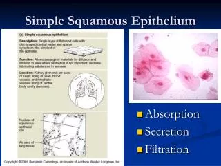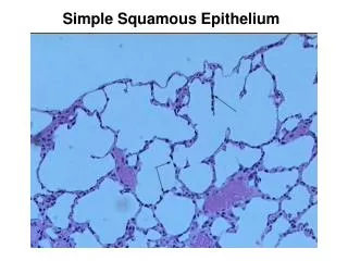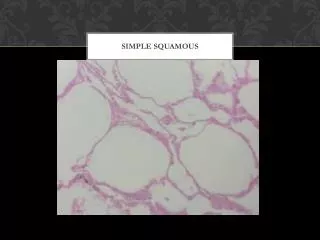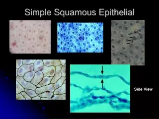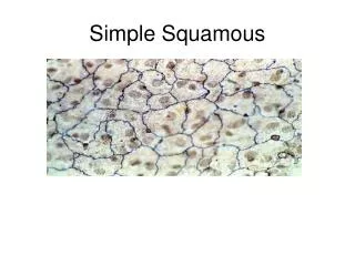
Simple Squamous Epithelium
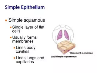
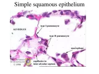
Simple Squamous Epithelium. alveoli and capillaries of lungs where gas exchange occurs kidney glomerulus and tubules where filtration and diffusion processes form urine capillaries where diffusion and osmosis occur ventral body cavities as mesothelium of serous membranes

Simple Squamous Epithelium
E N D
Presentation Transcript
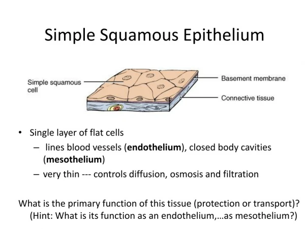
Simple Squamous Epithelium alveoli and capillaries of lungs where gas exchange occurs kidney glomerulus and tubules where filtration and diffusion processes form urine capillaries where diffusion and osmosis occur ventral body cavities as mesothelium of serous membranes all vessels and the heart as endothelium
Simple Cuboidal Epithelium • secretory portions exocrine and endocrine glands • ducts of many exocrine glands • kidney tubules
Simple Columnar Epithelium • ducts of exocrine glands • larger tubules or collecting ducts of the kidney • stomach, small intestine, and large intestine • smaller respiratory tubes or bronchioles • fallopian tubes • goblet cells
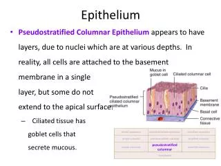
Pseudostratified epithelium • Nasal Mucosa
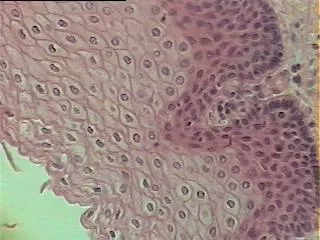
Stratified Epithelium“Squamous” • Keratinized stratified squamous epithelium is found only in the skin! • Thick skin has many layers of these dead cells cemented together. • Thin skin has fewer layers of living and dead cells but same structure.
Non Keratinized • the oral cavity • esophagus to the stomach junction • anus and rectum • vagina and cervix
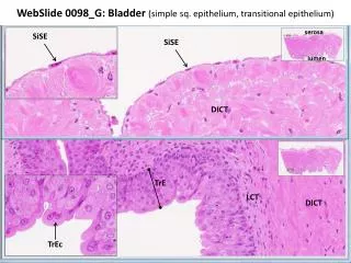
Transitional • Transitional epithelium is only found in the urinary tract! • Transitional epithelium in the bladder! • Transitional epithelium in the urethra!
Stratified Cuboidal and Columnar epithelium • In certain ducts and along transition zones in body tracts, stratified columnar and cuboidal epithelia can occur. As epithelial types, both are uncommon. Basal cells are typically cuboidal with surface cells either columnar or cuboidal in appearance. These types can be found in the larger ducts of various glands, including the pancreas, salivary, and sweat glands. If you are viewing an epithelium and it consists of more than 3-4 layers of cells, it will not be one of these types. Stratified squamous types and transitional are the only epithelia consisting of multiple cell layers.
Glands • Cells that secrete products via the merocrine method form membrane-bound secretory vesicles internal to the cell. These are moved to the apical surface where the vesicles coalesce with the membrane on the apical surface to release the product. Most glands release their products in this way.
Apocrine • In those glands that release product via the apocrine method, the apical portions of cells are pinched off and lost during the secretory process. This results in a secretory product that contains a variety of molecular components including those of the membrane. Mammary glands release their products in this manner.
Holocrine • The third type of secretory release, holocrine, involves death of the cell. The secretory cell is released and as it breaks apart, the contents of the cell become the secretory product. This mode of secretion results in the most complex secretory product. Some sweat glands located in the axillae, pubic areas, and around the areoli of the breasts release their products in this manner. Sebaceous glands also are of this type.
Endocrine • Endocrine glands are the hormone producing structures of the body. Some, like the thyroid are large and obvious. Others, for instance the islet cells of the pancreas, are small islands of endocrine cells embedded within the larger exocrine portion of this organ. • In lacking ducts, endocrine cells release their secretory products into the interstitial spaces around the cells. The hormones diffuse into nearby capillaries and are then carried to all parts of the body. Only when the hormones encounter a "target organ" do they exert an effect.
Connective Tissue • Connective tissues are the “glue” of the body. They connect parts to other parts • Fluid – lymph and blood • Connective Tissue Proper – loose and dense • Supportive Connective Tissue – bone and cartilage
CT Proper – Loose areolar • Connective tissue proper contains gel and fibers and the cells that produce them…remember…gels and fibers. What changes between tissue types are how much of each and the composition of each. • As the name implies, loose connective tissue consists of a loosely woven mix of fibers, cells, and ground substance. Areolar, a more technical name used for this tissue type means "spaces". Loose connective tissue therefore possesses randomly arranged protein fibers with abundant intercellular spaces. Scattered within the spaces are 7 cell types worth mentioning for their physiological roles:
Adipose with sweat gland Sweat gland
Reticular Connective - loose Although present as the supportive tissue of lymph nodes, glands, organs, and bone marrow, reticular connective tissue is not that obvious. Small, branching, collagen fibers that form the reticular connective tissue are usually hidden from view by the numerous lymphatic, epithelial, or bone marrow cells anchored to them. The stroma, or supporting network of reticular fibers is best seen with special stains.
Dense Connective Tissue – (fibrous type) tendon Notice the nuclei of fibroblasts squeezed and flattened in rows between the collagen fibers. Edges of these fibers are difficult to resolve and the elongate nuclei can be mistaken for those of smooth muscle. However, the "box car" arrangments of nuclei between the fibers is a key characteristic for dense, regular fibrous connective tissue.
DRCT (elastic type) In this side by side comparison of fibrous and elastic tendons cut in cross-section, the relative sizes of fibers is compared. Notice how large and robust the collagen fibers are compared to the smaller elastic types. Nuclei of fibroblasts visible along the edges of fibers are a good indicator these are tendons in cross-section. Elastic type Fibrous type
Irregular CT - collagen • in the dermis and hypodermis(thick skin) • in walls of organs forming the submucosa layer • in periosteum, perichondreum, perineurium(special stain), and perimysium • in the articular capsules of synovial joints
Osseous Tissue Osteon – cell which produces bone matrix
Hyaline cartilage • Hyaline Cartilage • The type of protein fiber embedded within the matrix of cartilage determines the cartilage type. In hyaline cartilage protein fibers are large and predominantly collagen. The optical density of these fibers is the same as the ground substance surrounding them and as a result, they are not visible within the extracellular matrix. Hyaline cartilage subsequently appears as a very uniform, glossy type tissue with evenly dispersed chondrocytes in lacunae. Typically, perichondrium is found around hyaline cartilage.