Download
1 / 15
160 likes | 463 Views
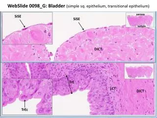
WebSlide 0098_G: Bladder (simple sq. epithelium, transitional epithelium). serosa. SiSE. SiSE. lumen. DICT. TrE. LCT. DICT. TrEc. WebSlide 0038_G: Submandibular gland (simple cuboidal epithelium lining secretory ducts). ducts in longitudinal section. ducts in cross section.

E N D
WebSlide 0098_G: Bladder (simple sq. epithelium, transitional epithelium) serosa SiSE SiSE lumen DICT TrE LCT DICT TrEc
WebSlide 0038_G: Submandibular gland (simple cuboidal epithelium lining secretory ducts) ducts in longitudinal section ducts in cross section
WebSlide 0032_G: Ileum (simple columnar epithelium) serosa LCT lumen jc bb epith gc gc gc LCT epith LCT
WebSlide 0051_G: Uterine Tube (simple columnar ciliated epithelium) lumen sm basal bodies cilia ncc epith
WebSlide0078A_G: esophagus (stratified sq. non-keratinized epithelium) basal cells surface squamous cells
WebSlide0065_G: skin, foot (stratified sq. keratinized epithelium) sweat gland duct basal cell layer “stratum lucidum” (lipid rich, so doesn’t stain very well) “stratum corneum” (outermost surface squamous cells) (note ABSENCE of nuclei in the stratum lucidum and corneum)
WebSlide0008_G: trachea (pseudostratified ciliated columnar epithelium) tracheal gland secretory duct basal cells basement membrane ciliated columnar epithelial cell (note height of cilia) goblet cell (note ABSENCE of cilia)
Slide UMich #30: abdominal mesentery examples of simple squamous epithelium: mesothelium endothelium
Slide UMich 9N-1: Kidney(simple cuboidal epithelium, coiled tubes in various planes of section)
Slide UMich 9N-1: Kidney(simple cuboidal epithelium, tubes mostly in longitudinal section)
Slide UMich 153: Esophagus (stratified squamous non-keratinized epithelium) GL(sce) adventitia LCT lumen duct duct DICT LCT artificial separation SM DICT StSE lumen StSEc
Slide UMich 106: Thick Skin(stratified squamous keratinized epithelium) Deep fascia connective tissue (dermis) blood vessel Superficial Fascia epithelium (epidermis) keratinized layer
Slide UMich 40: trachea (pseudostratified columnar ciliated epithelium with goblet cells) Cilia Basal bodies Goblet cell Basement membrane