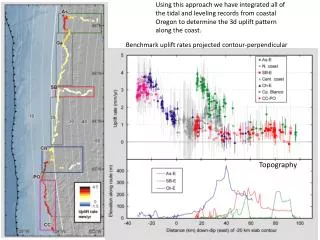
Brain Topography
Brain Topography. Cerebrum. Cerebrum. Cerebellum. Cerebrum. The largest division of the forebrain . It is divided into two hemispheres, each of which is divided into four lobes. Corpus callosum.

Brain Topography
E N D
Presentation Transcript
Cerebrum Cerebrum Cerebellum Cerebrum The largest division of the forebrain. It is divided into two hemispheres, each of which is divided into four lobes.
Corpus callosum • Divided into two halves, the cerebral hemispheres, whichare separated by a deep Median longitudinal fissure which lodges a dural fold called falxcerebri. • In the depth of the fissure, the hemispheres are connected by a bundle of fibers called the corpus callosum. Right hemisphere Left hemisphere Median Longitudinal fissure
The structure of cerebral hemispheres includes: Superficial layer of grey matter, the cerebral cortex. Deeper to the cortex, axons running to and from the cells of the cortex form an extensive mass of white matter (WM). Burried within the white matter lie a number of nuclear masses (caudate, putamen, globuspallidus) collectively known as the basal ganglia. The cavity of hemisphere is called the lateral ventricle. Cortex Basal ganglia WM Lateral ventricle
Cerebral Cortex CerebralCortex Cerebral Cortex The outermost layer of gray matter making up the superficial aspect of the cerebrum.
Cerebral Features • Gyri– Elevated ridges “winding” around the brain. • Sulci– Small grooves dividing the gyri • Central Sulcus – Divides the Frontal Lobe from the Parietal Lobe Highly convoluted arrangement. This arrangement maximize the surface area of the cerebral cortex (about 70% is hidden within the depths of sulci). • Fissures– Deep grooves, generally dividing large regions/lobes of the brain • Longitudinal Fissure – Divides the two Cerebral Hemispheres • Transverse Fissure – Separates the Cerebrum from the Cerebellum • Sylvian/Lateral Fissure – Divides the Temporal Lobe from the Frontal and Parietal Lobes
Gyri (ridge) Sulci (groove) Fissure (deep groove)
Specific Sulci/Fissures: Central Sulcus Longitudinal Fissure Sylvian/Lateral Fissure Transverse Fissure
Three sulci, consistent in their position (central, lateral & parieto-occipital) are used to divide each hemisphere into lobes. • Each hemisphere is divided into FOUR lobes (named after overlying bones). reception and evaluation of sensory information motor function, motivation, aggression, smell and mood visual processing smell, hearing, memory and abstract thought
Frontal Parietal Occipital Temporal Lobes of the Brain (4) * Note: Occasionally, the Insula is considered the fifth lobe. It is located deep to the Temporal Lobe.
The Frontal Lobe of the brain is located deep to the Frontal Bone of the skull. Lobes of the Brain - Frontal • It plays an integral role in the following functions/actions: • - Memory Formation • - Emotions • - Decision Making/Reasoning • Personality • In addition to its major role in motor functions
Frontal lobe • Precentralgyrus. • Superior & inferior frontal sulci divide the lobe into superior, middle & inferior frontal gyri.
Frontal Lobe Premotor cortex: Located in the region immediately anterior to the precentralgyrus(Brodmann’s area 6). Primary motor cortex: Located in precentralgyrus(Brodmann area 4). Prefrontal cortex: Extensive region of the frontal lobe anterior to premotor area. Broca’s (motor speech) area: Located in the inferior frontal gyrus of the dominant hemisphere, usually left (Brodmann’s area 44 & 45). Frontal eye field: Located in the middle frontal gyrus immediately in front of motor cortex (Brodmann’s area 8).
Gyri of the Frontal Lobe Lateral Surface PrecentralGyrus --- primary motor area: Superior Frontal Gyrus Middle Frontal Gyrus Inferior Frontal Gyrus Pars Opercularis Pars Triangularis Pars Orbitalis Medial Surface Medial Frontal Gyrus Paracentral Lobule Basal Surface Rectus Gyrus Orbital Gyrus Inferior Frontal Gyrus ] Broca’s area (dominant hemisphere)
Gyri of • the Cerebral Cortex • (basal surface) Basal Surface Rectus Gyrus Orbital Gyrus Inferior Frontal Gyrus
Gyri of the Cerebral Cortex (Medial Surface) Medial Surface Medial Frontal Gyrus Paracentral Lobule
The Parietal Lobe of the brain is located deep to the Parietal Bone of the skull. Lobes of the Brain - Parietal Lobe • It plays a major role in the following functions/actions: - Senses and integrates sensation(s) • Spatial awareness and perception • (Proprioception - Awareness of body/ body parts in space and in relation to each other)
Parietal Lobe • Parietal lobe: • Postcentralgyrus. • Intraparietal sulcus dividing the lobe into superior & inferior parietal lobules.
Primary Somatosensory Cortex/ PostcentralGyrus Somatosensory Association Cortex Primary Gustatory Cortex
Parietal Lobe • Primary Somatosensory Cortex (PostcentralGyrus)– Site involved with processing of tactile and proprioceptive information. • Somatosensory Association Cortex - Assists with the integration and interpretation of sensations relative to body position and orientation in space. May assist with visuo-motor coordination. • Primary Gustatory Cortex– Primary site involved with the interpretation of the sensation of Taste.
Parietal lobe Primary somatosensory cortex: located in postcentralgyrus(Brodmann’s area 1, 2, 3). Parietal association cortex: located posterior to primary somatosensory cortex.
Gyri of the Parietal Lobe • Lateral Surface • PostcentralGyrus---- • Superior Parietal Lobule • Inferior Parietal Lobule ---- Wernicke’s area ?? SupramarginalGyrus Angular Gyrus • Medial Surface • Paracentral Lobule • Precuneus
Lobes of the Brain – Occipital Lobe • The Occipital Lobe of the Brain is located deep to the Occipital Bone of the Skull. • Its primary function is the processing, integration, interpretation, etc. of VISION and visual stimuli.
Primary Visual Cortex– This is the primary area of the brain responsible for sight -recognition of size, color, light, motion, dimensions, etc. located on the medial surface of the hemisphere, in the gyri surrounding the calcarine sulcus (Brodmann’s area 17). Occipital Lobe • Visual Association Area– Interprets information acquired through the primary visual cortex. located around the primary visual cortex.
Primary Visual Cortex Visual Association Area
Gyri of the Occipital Lobe Lateral Surface: Lateral Occipital Gyrus Superior Occipital Gyrus Inferior Occipital Gyrus Medial Surface Cuneus Lingual Gyrus Basal Surface Lingual Gyrus OccipitotemporalGyrus
Lobes of the Brain – Temporal Lobe • The Temporal Lobes are located on the sides of the brain, deep to the Temporal Bones of the skull. • They play an integral role in the following functions: • Hearing • Organization/Comprehension of language • Information Retrieval (Memory and Memory Formation)
Temporal lobe: Superior & inferior temporal sulci giving rise to superior, middle & inferior temporal gyri. Insula: the gyri in the depth of lateral fissure, covered by parts of frontal, parietal & temporal lobes called the opercula (removed in lower picture.). Superior, middle & inferior temporal gyri sts its insula
Primary Auditory Cortex– Responsible for hearing located in the superior surface of the superior temporal gyrus(Brodmann’s area 41, 42) Temporal Lobe • Primary Olfactory Cortex– Interprets the sense of smell once it reaches the cortex via the olfactory bulbs. (Not visible on the superficial cortex) Auditory association cortex: located immediately around the primary auditory cortex (also includes Wernick’s area) • Wernicke’s Area– Language comprehension. Located on the Left Temporal Lobe.
Primary Auditory Cortex Wernike’s Area Primary Olfactory Cortex (Deep) Conducted from Olfactory Bulb
Temporal Lobe • Parahippocampalgyrus: • located in the inferomedial part of temporal lobe. • Deep to this gyrus lies the hippocampus and the amygdala, which are parts of limbic system
Language Area • Organized around the lateral fissure. • Broca’s area: concerned with expressive aspects of language. • Wernick’s area: responsible for comprehension of the spoken words. • Nearby regions of temporal lobe and parietal lobe (angular gyrus & supramarginalgyrusof the inferior parietal lobule) are important in naming, reading, writing, and calculation.
Arcuate Fasciculus • Arcuate Fasciculus- A white matter tract that connects Broca’s Area and Wernicke’s Area through the Temporal, Parietal and Frontal Lobes. Allows for coordinated, comprehensible speech. Damage may result in: - Conduction Aphasia - Where auditory comprehension and speech articulation are preserved, but people find it difficult to repeat heard speech.
Gyri of the Temporal Lobe Lateral Surface Superior Temporal Gyrus Middle Temporal Gyrus Inferior Temporal Gyrus Basal Surface Lingual Gyrus OccipitotemporalGyrus Medial OccipitotemporalGyrus Lateral Occipitotemporal
Somatosensory cortex (Somesthetic sensation and proprioception) Supplementary motor area (programming of complex movement) Primary motor cortex (Voluntary movement) Premotor cortex (coordination of complex movements) Posterior parietal cortex (integration of somatosensory and visual input) Central sulcus Prefrontal association cortex (planning for voluntary activity; decision making; personality traits) Parietal lobe Wernicke’s area (speech understanding) Frontal lobe Parietal-temporal-occipital association cortex (integraton of all sensory input- imp in language) Broca’s area (speech formation) Primary auditory cortex Occipital lobe Limbic association cortex (motivation, emotion, memory) Temporal lobe Primary visual cortex
Lobes and Structures of the Brain A. Central Sulcus B. Frontal Lobe C. Sylvian/Lateral Fissure A. (groove) G. D. Temporal Lobe B. F. E. Transverse Fissure F. Occipital Lobe C. (groove) G. Parietal Lobe E. D. (groove)
Hemispheric Dominance • The localization of speech centers & mathematical ability is the criterion for defining the dominant cerebral hemisphere. • In 96% of normal right-handedindividuals and 70% of normal left-handed individuals, the left hemisphere contains the language centers. These are left hemisphere dominant. • Cerebral dominance becomes established during the first few years after birth. Verbal Memory Shape Memory Hemispheres communicate via the corpus callosum
White Matter Underlies the cortex and contains: • Nerve fibers, Neuroglia cells & Blood vessels. • The nerve fibers run in different directions and originate, terminate or sometimes both, within the cortex.
Depending on their origin & termination, the nerve fibers in the cerebral white matter are classified into three types: Association, Commissural & Projection Association fibers: Unite different parts of the same hemisphere Commissural fibers: Connect the corresponding regions of the two hemispheres • Projection fibers: Consisting of • Afferent fibers conveying impulses to the cerebral cortex. • Efferent fibersconveying impulses away from the cortex.
Association Fibers Short association fibers • Unite different parts of the same hemisphere. • Are of two kinds: • Those connecting adjacent gyri, short association fibers • Those connecting more distant parts, long association fibers. Long association fibers
Uncinate fasciculus: connects frontal to temporal lobe Superior longitudinal fasciculus: connects the frontal, occipital, parietal, and temporal lobes Arcuate fasciculus: connect gyri in frontal to temporal lobes Inferior longitudinal fasciculus: connects occipital to temporal pole Long Association Fibers 2 5 3 1 4 5. Cingulum: connects frontal & parietal lobes to the para-hippocampal gyrus and adjacent temporal gyri
Commissural Fibers • Connect the corresponding regions of the two hemispheres. • Include: • Corpus callosum. • Anterior commissure. • Hippocampal commissure (commissure of fornix). • Posterior commissure.
Corpus Callosum Anterior forceps • Connects the corresponding regions of the two hemispheres except the temporal lobes, that are connected by anterior commissure • It is shorter craniocaudally than is the hemisphere • The callosal fibers linking the frontal poles curve forward forming anterior forceps (forceps minor) • The callosal fibers linking the occipital poles curve backward forming posterior forceps (forceps major) F CC P O Posterior forceps
Parts of Corpus Callosum Body Genu Splenium Rostrum
Posterior Commissure: connects the left and right midbrain. Important in the bilateral pupillary reflex • Anterior commissure: connects the inferior and middle temporal gyri & the olfactory regions of the two hemispheres • Hippocampal Commissure: connects the two hippocampi with each other