Download

1 / 72
750 likes | 1k Views
Role of CT in acute pancreatitis. Dr. Ahmed Refaey. Consultant radiologist Riyadh Military Hospital. MBBCh , MS, FRCR . Normal CT anatomy of the upper abdomen. Anterior pararenal space. Pancreas is located in the anterior pararenal space Head adjacent to duodenum

E N D
Role of CT in acute pancreatitis Dr. Ahmed Refaey Consultant radiologist Riyadh Military Hospital MBBCh, MS, FRCR
Pancreas is located in the anterior pararenal space Head adjacent to duodenum Tail extending toward spleen Splenic vein posterior to body and tail Normal Anatomy by CT
Normal Morphology by CT • No capsule • AP dimensions • Head 2-2.5 cm • Body and tail 1-2 cm • Pancreatic duct • Maximal diameter 3 mm in adults (5 mm in elderly)
Evaluation of Acute Pancreatitis • Contrast-enhanced CT is imaging modality of choice • Oral and IV contrast differentiate pancreatic tissue from adjacent blood vessels and duodenum
There is no additional value of an early CT (within 72 hours) in patients with acute pancreatitis. • The diagnosis is usually made on clinical and laboratory findings. • An early CT may be misleading concerning the severity of the pancreatitis, since it can underestimate the presence and amount of necrosis.
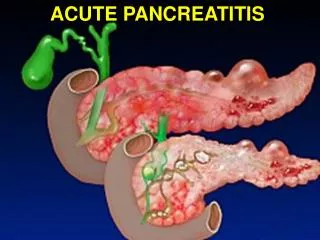
Pathophysiology • Activated pancreatic enzymes escaping the ductal system and auto digesting the pancreas and adjacent structures ( mainly amylase, lipase & trypsin ). • Lack of capsule facilitates spread
Acute pancreatitis • Mild acute pancreatitis ----- 80 % * edematous ( interstitial ) * exudative • Severe acute pancreatitis ----20 %
Mild acute pancreatitis - run a mild course without development of multiple organ failure - improvement within 3 days following conservative therapy with gradual decrease of elevated enzymes. - has a mortality rate of < 1%
Severe acute pancreatitis “ necrotizing “ - run a serious clinical course with pancreatic necrosis and the development of multiple organ failure - of these, 60% of pancreatic necrosis remain sterile , while 40% becomes infected - this last category ( infected necrosis ) , has the highest mortality rate ( 25-70%)
Mild acute pancreatitis • Acute edematous “interstitial” pancreatitis • Acute exudative pancreatitis
Acute edematous “interstitial” pancreatitis • Edematous pancreas with/without peripancreatic fat stranding. • No collections or necrosis
Acute exudative pancreatitis • an intermediate form of pancreatitis without pancreatic necrosis with an intermediate clinical course. • This is called extrapancreatic necrosis (EXPN)
Avoid early drainage of collections and avoid introducing infection! • 50% of these collections show spontaneous regression The other 50% either remain stable ( pseudocyt ) or develop infection ( abscess ) .
Peripancreatic fluid 50% spontaneous regression 50% stable sterile ( pseudocyst) infection ( asbscess )
Severe pancreatitis “ necrotizing pancreatitis” occurs in 20% of patients. * partial necrotizing pancreatitis * total necrotizing pancreatitis
Partial necrotizing Total necrotizing • Delayed or no response to conservative therapy • Delayed or no normalization of enzymes • Mortality : 30 – 75 % • Deterioration under conservative therapy • Mortality : 100 % - 40% by 2nd day - 75% by 5th day - 100% by 10th day
Central gland necrosis • Subtype of necrotizing pancreatitis. • Necrosis between the pancreatic head and tail and is nearly always associated with disruption of the pancreatic duct. • This leads to persistent collections as the viable pancreatic tail continues to secrete pancreatic juices. • These collections react poorly to endoscopic or percutaneousdrainage. • Definitive treatment often requires distal pancreatectomy.
An early CT may be misleading concerning the severity of the pancreatitis, since it can underestimate the presence and amount of necrosis.
Mortality • Early mortality in acute pancreatitis is the result of the systemic inflammatory response with multiple organ failure.Late mortality is the result of infection of pancreatic necrosis and peripancreatic fluid collections which results in sepsis and is seen in more than 50% of deaths.
CT Severity Index • It is critical to identify patients who are at high risk for severe disease, since they require close monitoring and possible intervention
Balthazar et al constructed a CT severity index (CTSI) for acute pancreatitis that combines the grade of pancreatitis (A-E) with the extent of pancreatic necrosis.
CT Severity Index • 1- 3 …………….. Mild • 4-6 …………….. Moderate • 7-10 ……………. Severe