Download

1 / 64
760 likes | 1.49k Views
Acute Pancreatitis. Resident Conference October 5, 2004 Rachel Dunagin, MD. Background. Acute Inflammatory process of pancreatic parenchyma A stimulus leads to release of activated digestive enzymes from acinar cells into interstitium Results in autodigestion of pancreas and adjacent tissue

E N D
Acute Pancreatitis Resident Conference October 5, 2004 Rachel Dunagin, MD
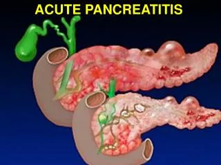
Background • Acute Inflammatory process of pancreatic parenchyma • A stimulus leads to release of activated digestive enzymes from acinar cells into interstitium • Results in autodigestion of pancreas and adjacent tissue • Activated inflammatory mediators convert a localized inflammatory response into a systemic inflammatory process, resulting in increased tissue and vascular permeability • End result: hypovolemia, shock, ARDS, multisystem organ failure • Majority: mild self-limited course; 15-25% severe, complicated course; 5% mortality
Gallstone (35%) Microlithiasis Alcoholism (30%) Medication Trauma/Sphincter of Oddi Dysfunction Infection Duodenal Diverticula Hypercalcemia Vascular Abnormalities Post-operative Neoplasm Pancreas Divisum Autoimmune Hereditary Idiopathic (30%) Hypertriglyceridemia Cystic Fibrosis Etiology
Etiology: Gallstones • ½ of all cases of acute pancreatitis • 3-8% patients with symptomatic cholelithiasis develop pancreatitis • Older women • 80% patients previously thought to have idiopathic etiology are due to microlithiasis, tiny gallstones, and biliary sludge • Mechanism unclear – pancreatic ductal obstruction does not full explain pathogenesis
Etiology: Alcohol • Interferes with normal process of pancreatic secretion • Excessive deposition of GP-2 (protein involved in maintaining normal pancreatic secretion) leads to ductal obstruction • Daily consumption 80g alcohol over 5-15 years before first attack of alcoholic pancreatitis occurs • Acute attacks 1-3 days after drinking
Etiology: Hypertriglyceridemia • >1000mg/dL • 50% with hyperTG have falsely normal amylase level due to interference of lipemic specimen with assay • Therefore, must dilute serum to get accurate serum amylase level
Etiology: Medications • Uncommon cause • Azathioprine, 6-mercaptopurine and ddI have 5-10% risk of acute pancreatitis • >100 meds implicated
Azathioprine Asacol Cytosine arabinoside Estrogens Norethindrone/mestr-anol Isoniazid 6-mercaptopurine Metronidazole Pentamidine Tetracycline Trimethoprim/sulfamethoxazole (TMP/SMX) Valproic Acid Definitely Cause Pancreatitis
Implicated Agents • Industrial chemicals (Parathion) • Scorpion venom
Viral Infections • Mumps – most common in adults • Coxsackie-B • Epstein-Barr • Rubella • Influenza A • Varicella • Hepatitis A, B, C, E
AIDS and Pancreatitis • 10% with AIDS develop acute pancreatitis • Asymptomatic pancreatic lesions in 30-50% of autopsy cases • Common infectious etiologies: CMV, MAC, Crypto, M. TB, Toxoplasma • Common medication etiologies: pentamidine, ddI, ddC and TMP/SMX
Rare Etiologies • SLE • Polyarteritis Nodosum • Autoimmune pancreatitis • Fungal infections • Bacterial infections: Legionella, Brucellosis • Ampullary tumors • Metastatic tumors (breast, lung) • Pancreas Divisum
Pancreatic Divisum • Debatable cause • Represents 5-7% of population per autopsy and ERCP studies • Normal Pancreatic Development: • Dorsal and ventral pancreatic buds fuse during 8th week gestation to form main pancreatic duct; drains through major papilla. • Small duct often persists between the main pancreatic duct and the minor papilla
Pancreatic Divisum • Embryonic dorsal and ventral ducts fail to migrate and fuse abnormally. • Two noncommunicating duct systems: • Inferior portion of head of pancreas drained by rudimentary ventral duct through the major papilla. • Remainder of pancreas drained by dorsal duct through the minor papilla.
Pancreatic Divisum:Hypothesis of pancreatitis etiology • First proposed in 1977 • Increased resistance to flow through a small minor papilla. • If so, would reason that recurrent pancreatitis would stop after endoscopic minor papilla sphincterotomy, or insertion of endoscopic stent across the minor papilla. • Some disagree on basis that incidence of pancreas divisum is the same in patient with or without pancreatitis and 95% with pancreatic divisum do not develop pancreatitis • Other arguments against: pancreatitis develops in adulthood, not childhood in those with pancreatic divisum • Still remains controversial.
Diagnosis Clinical signs and symptoms, confirmed with lab/radiologic studies Symptoms: • Acute abdominal pain • Location: entire abdomen or localized in midepigastric, RUQ or left flank • Intensity: maximized within 10 – 20 min of acute attack • Quality: steady, moderate to severe, little relief with change of position, unbearable, refractory to narcotics • Radiation: band-like to the back • Anorexia, nausea, emesis • CNS manifestations: disorientation, hallucinations, agitation, or coma
Diagnosis Signs: Mild Pancreatitis – mild abdominal tenderness, no guarding Severe Pancreatitis - epigastric tenderness and guarding, rebound tenderness, abdominal distention (due to gastric ileus or dilatation of transverse colon) • Decreased or absent bowel sounds • May be mistaken for acute abdomen • If severe, extensive peripancreatic fat necrosis with hemorrhagic fluid within the peritoneum and/or retroperitoneum → Cullen’s sign, Grey-Turner’s sign • Palpable epigastric mass = pseudocyst or large inflammatory mass • Subcutaneous nodular fat necrosis, thrombophlebitis in legs, polyarthritis
Diagnosis • Due to third-space fluid loss and systemic toxicity • Pulse 100-150 • BP hypertensive in beginning, then hypotensive due to 3rd spacing and hypovolemia • Temp: initially normal, then increase to 101-103 within 1-3d • Tachypnea and shallow respirations
Nonspecific Lab Findings • Leukocytosis • Hyperglycemia • Hypocalcemia • LFTs: AST, ALT, AlkPhos, Bilirubin • Hypertriglyceridemia • Hypoxemia
Laboratory Testing • Serum Amylase • Most widely accepted • 2-3x upper limit of normal • Does not correlate with severity of disease • 30% of Alcoholics with acute pancreatitis have normal amylase levels • ? of using elevated lipase to amylase level to predict alcoholic acute pancreatitis • ALT 3x upper limit of normal + elevated amylase is highly sensitive for gallstone pancreatitis (95% PPV)
Laboratory Tests • Caution!! -- Other causes of elevated amylase level • Can be of pancreatic or salivary origin • Any inflammatory process of abdomen: • Perforated peptic ulcer • Intestinal obstruction • Cholecystitis • Generalized peritonitis • Mesenteric ischemia • Ruptured AAA/ectopic pregnancy • Renal failure • Macroamylasemia: due to benign change in peptide processing in Golgi (amylase is bound to immunoglobulin or abnormal serum protein), forms a large complex which is difficult to clear through the kidneys
Laboratory Testing • Serum Lipase • More sensitive and specific than amylase • Greater than 2-3x upper limit of normal • Does not correlate with severity of disease
Other Lab Tests • Pancreatitis associated protein (PAP), heat shock protein • Trypsinogen activation peptide (TAP), 5-amino acid peptide cleaved from trypsinogen to produce active trypsin • Non-specific Markers: CRP, neutrophil elastase, complement, TNF, IL6 • Methemalbumin level
Radiology • Sonography • Computer Tomography (CT) • Magnetic resonance imaging (MRI)
Ultrasound • Fails to completely to completely image the pancreas 2/2 overlying bowel gas • Superior to CT in visualizing gallbladder and biliary tree for gallstones • Negative US does not rule out gallstone as etiology because: 1. Not sensitive for common bile duct stones. 2. Only 50% of those with microlithiasis have + US. 3. View may be obstructed by ileus and underlying structures obscured by bowel gas.
A B Acute Pancreatitis. A. Transverse scan. B. Longitudinal scan. The head of the pancreas (H) is enlarged as revealed by the red arrowheads and decreased in echogenicity because of edema. The surrounding structures are superior mesenteric vein (v), superior mesenteric artery (a), abdominal aorta (A), and inferior vena cava (IVC).
Computer Tomography (CT) • Imaging modality of choice for diagnosis, determining severity, and identifying complications • Sensitivity: 90% for diagnosis of acute pancreatitis • Specificity: 98-100% • Not necessary for mild acute pancreatitis; however is useful to rule out other abdominal processes presenting with abdominal pain. • Mild disease: no abnormalities, diffuse enlargement of pancreas, loss of normally sharp border, homogenous attenuation, inflammatory stranding in peripancreatic fat and adjacent soft tissue, fluid collections, pseudocysts, pancreatic necrosis
Computer Tomography (CT) • Necrotizing pancreatitis: Non-enhancement of ≥ 1/3 of pancreas or >3cm of non-enhancement of the pancreas on dynamic, IV contrast-enhanced CT. If > 30% gland involved, sensitivity approaches 100%
Indications for CT • Severe acute pancreatitis (Ranson score ≥3 or APACHE II score ≥8) • Mild pancreatitis with no response to conservative management after 48-72 hours (confirm dx, re-assess severity, identify complications) • May repeat q7-10 day if no improvement or if deterioration.
Severity • Not predicted by degree of pain, etiology or serum amylase level. • CRP: > 120mg/L in pts with acute pancreatitis typically have necrotizing pancreatitis. • Phospholipase A2: elevated in severe disease, esp those who develop necrosis, ARDS, and shock • Urinary trypsinogen activation peptide: ≥ 10ng/mL has 100% PPV of severe pancreatitis • Peritoneal Lavage: any volume of peritoneal fluid with a dark color or recovery of at least 20mL of free intraperitoneal fluid = 33% mortality
Severity • Hematocrit ≥ 47% at admission or failure of admission hematocrit to decrease at 24hrs is strong predictor of development of pancreatic necrosis. • Compromised microcirculation of pancreatic parenchyma precipitated by fluid sequestration with intravascular volume depletion and hemoconcentration leads to development of necrotizing pancreatitis. • Therefore, intense hydration is important.
Severity • Ranson Criteria ≥ 3 • APACHE II score ≥ 8 • Hematocrit ≥ 44 • CT severity index ≥ 6 • 1992 Atlanta Symposium: evidence of organ failure or local complications
Ranson Criteria **Can not be applied fully for 48 hours **Poor predictor later in the disease **Single snapshot in time
APACHE II Criteria • Multivariate scoring system • 12 measurable variables: temperature, heart rate, respiratory rate, mean arterial BP, oxygenation, arterial pH, serum potassium, sodium and creatinine, hematocrit, WBC, and Glasgow Coma Scale • Account for premorbid state and age • Can be used throughout course of illness, but is complex and cumbersome
CT Severity Index A: Normal pancreas B: Enlargement of pancreas, heterogeneous enhancement without peripancreatic disease C: Pancreatic abnormalities with peripancreatic disease D: Small or single fluid collection E: 2 or > fluid collection or pancreatic abscess
Atlanta Symposium Criteria • 40 Internationally renowned experts on pancreatic disease met to define severity of pancreatitis • Defined based on outcome: organ failure and/or anatomic complications • Mild acute pancreatitis: minimal or no organ system dysfunction with complete and uneventful recovery; interstitial edema of parenchyma • Severe acute pancreatitis: evidence of life-threatening systemic complications or pancreatic collection.
Severe Acute Pancreatitis • Systemic Complications: • Shock: SBP <90mm Hg • Pulmonary Insufficiency: PaO2≤ 60mm Hg • Renal failure: Cr > 2mg/dL after rehydration • GI bleeding: > 500ml/24hr • DIC: platelets < 100,000/mm3, fibrinogen < 1g/L, fibrin degradation products > 80mug/mL • Hypocalcemia: < 7.5mg/dL
Severe Acute Pancreatitis • Pancreatic Collections • Pancreatic Necrosis: diffuse or focal areas of nonviable pancreatic parenchyma • Pancreatic Abscess: well-circumscribed collection of pus containing little or no necrotic tissue • Acute Pseudocyst: collection of enzyme-rich, pancreatic fluid enclosed by a wall of fibrous tissue
Treatment • Goals: halt progression of local disease and prevent remote organ failure • Nutritional Support: • Pancreatitis is catabolic state • Benefit of pancreatic “rest” by limiting oral intake is unproven, however is widely used • Evidence that early enteral nutrition is safe • Nasojejunal feeding limits pancreatic secretion • Preferable to oral or nasogastric feeding
Treatment • Fluids • IV hydration, aggressive (300-500ml.hr) especially in the early phase of illness with goal hemodilution to Hct 30% • May need NG tube (if persistent nausea and vomiting or ileus), Foley catheter and central line or Swan-Ganz catheter to monitor hydration status • Analgesics • Adequate pain control is essential • 50-100mg Meperidine (Demerol) IV q3-4hr • Hydromorphone (Dilaudid) PCA • Avoid Morphine b/c it increases sphincter of Oddi tone and increases serum amylase • ERCP • If severe acute gallstone pancreatitits or ascending cholangitis is indicated, then early ERCP with sphincterotomy and stone extraction is indicated. • Is not to be used in mild acute pancreatitis
Pancreatic Infection:A Word on Antibiotics • Antibiotics – there is no role for routine use • Indicated if severe attack with necrosis of pancreas • Typical organisms: from gut = E coli, Pseudomonas, Klebsiella, Enterococcus • Treatment: selective decontamination of gut with oral nonabsorbable antibiotics, systemic antibiotics, and enteral feedings to avoid catheter-related infections. • Imipenem/cilastin penetrate pancreatic parenchyma and reduces incidence of intra-abdominal infection. • Unfortunately, there is a tendency for fungal superinfection to develop later in clinical course. • Other options: 3rd gen cephalosporin, piperacillin, mezlocillin, fluoroquinolones and metronidazole