Download
1 / 41
450 likes | 715 Views
Role of Imaging Methods in Diagnosis of Acute Pancreatitis. Válek V. Radiologická klinika, FN Brno a LF MU v Brně. New Classification: Acute Pancreatitis.

E N D
Role of Imaging Methods in Diagnosis of Acute Pancreatitis Válek V. Radiologická klinika, FN Brno a LF MU v Brně
New Classification: AcutePancreatitis 2007 revision of Atlanta classification and definitionsof collections associated with acute pancreatitis by Acute Pancreatitis Working Group.
RevisedDefinitions: AcutePancreatitis • Patients who present with two of the following three manifestations are diagnosed as having acute pancreatitis: • Abdominal pain suggestive of pancreatic origin • Serum amylase, lipase ≥ 3 times normal • Characteristic findings on CECT • Disease Severity • First week based on clinical parameters • Thereafter on morphologic parameters (CECT) • Acute Pancreatitis Classification Working Group May 2007
Why CECT?? CT study is the most appropriate procedure to confirm image findings of acute pancreatitis. The evaluation of severity (CTSI) is basic, because it is strictly correlated to the prognosis of the patient. Early scanning for the prediction of severity is limited because the full extent of pancreatic necrosis may not develop within the first 48 hour of presentation.Dynamic CECThave80 – 90 % accurate for necrosis detection. Balthazar Radiology 1994; 193:297-306 Balthazar Radiology 2002; 223:603-613
CTSI Accuracy of CTSI for predicting pancreatitis severity: CTSI > 3 Sensitivity– 11/13 Specificity– 41/42 PPV – 11/12 NPV – 52/55 Accuracy – 52/55 Gurleyik et al J Pancreas 2005; 6:562-567
Acutepancreatitis The MR has actually a secondary role for the diagnosis, but it represents, first instance method in patients with adverse reaction to contrast medium. Standard MRI techniques including T1-weighted and T2-weighted fat-suppressed imaging sequences together with contrast-enhanced imaging.
CECT vesus MR Non-severe acutepancreatitis
Acutepancreatitis The ultrasound can be used as first instance method in patient with clinical suspect of acute pancreatitis. Contrast-enhanced ultrasound is a relatively new technique, currently used for liver tumors diagnosis. Contrast-enhanced ultrasound might represent a valuable additional imaging method to contrast CT for selected cases. Pulse inversion harmonic imaging allows the assessment of the necrotic areas in acute pancreatitis.
Old term: Mildacutepancreatitis New Term: Non-severe acutepancreatitis Histology: interstitial edema, micronecrosis Clinical course no MSOF (Multisystem organ failure) improve in 48 – 72 hr Imaging None → CECT US to evaluategallstone
Old term: Mildacutepancreatitis New Term: Non-severe acutepancreatitis
Old term: Severe acute pancreatitis New Term: Severe Acute Pancreatitis
5/1 17/1 6/4 26/12
New Classification: Necrosis: Based on CECTor MR • One or more focal areas of nonenhancing pancreatic parenchyma • Focal parenchymal low signal in T1 FS unenhanced sequence • Typically accompanied by gross peripancreatic fat necrosis • MR more heterogeneous than CT • May not be apparent up to 48 hours after onset • Potential pitfalls: • Apparent diminished enhancement value in patients with normal fatty infiltration • Patients with diffuse parenchymal edema in less severe, interstitial pancreatitis • Small intrapancreatic focal fluid collections
CECT vesus MR: Necrosis
New Classification: Fluid collections: CECT or MRI Enzyme rich pancreatic juice Acute peripancreatic fluid collection (APFC) (occurwithin 48 hrs in 30 – 50 %, majority remain sterile, resolves spontaneously within 2-4 weeks) Post necrotic pancreatic fluid collections (PNPFC) (fluid and necrotic contents –fat, initial necrosis → liquefactive necrosis Walled off pancreatic necrosis (WOPN) = late stage of PNPFC Pancreatic pseudocyst (4 weeks, contain NO necrosis) Pancreatic abscess = infected pseudocyst (dif.dg. Infected PNPFC and infected WOPN
Old term: Acute Fluid Collection New Term: Post Necrotic Pancreatic Colections (PNPC)
Old term: Pancreatic Pseudocyst New Term: Pancreatic Pseudocyst Well circumscribed, thin walled, homogeneous low attenuation collection of pancreatic juice. Requires 4 weeks, contains no necrosis Resolves spontaneously within 6 weeks 40%, 80% if <6cm Ductal communication – important for management Noninfected or infected (suppurative)
Old term: Pancreatic Absces New Term: Infected Pseudocyst Wall thicker, more irregular than the well delineated thin wall circumscribed collection of pus near pancreaswith homogeneously low attenuation center. Requires 4 weeks to form Contains little or no necrosis Should be differentiated from infected PNPC and infected WON
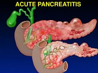
Role of Imaging Methods in Diagnosis of Acute Pancreatitis Early diagnosis Staging (grading) + classification Differential diagnosis Follow-up (treatment)
CECT = gold standard Reliable method Widely available Show local extend of inflammation/necrosis and occurrence of complications Balthazar CTSI – based on grade of pancreatitis and amount of glandular necrosis CTSI of 7 – 10 associated with 92% morbidity, 17% mortality Pts with CTSI ≤3 can safely be discharged from ICU Vriens et all, J.Am.Coll.Surgery, 2005, 2001, 491 – 502 Che et al, Eur.J.Radiol., 2006, 57, 102 – 107 Leung et al, World J. Gastroenterol., 2005, 11, 6049 - 6052
MRI = secondchoice Geographic areas of altered T1 signal correspond to areas of pancreatic necrosis depicted on CECT High signal intensity on unenhanced T1 FS SGE images corresponds to hemorrhage and correlates with severity. Sensitivity of MRI for acute pancreatitis may surpass CT (minor peripanc. inflamm) on T2 FS images MRSI =CTSI using dynamic 3DT1 SGE gado enhanced images, with added benefit of PD duct eval. Using secretin MRCP. PD duct evaluation using secretin MRCP Lecesne et al Radiology 1999; 211:727-735 PamuklarMagnResonClin NA 2005; 13:313-330 Arvanitakis Gastroenterology 2004; 126:715-723
Diagnosis?? Pancreaticatrophy Acutepancreatitis VonWildelmuthdisease Pancreaticagenesis Cysticfibrosis
Cysticfibrosis Cystic fibrosis is a condition with autosomal recessive inheritance in which there are defects of serous and mucous secretion involving multiple organs; 85 % of patients have severe exocrine pancreatic insufficiency and steatorrhoea. Obstruction of the main pancreatic duct and side branches by inspissated secretions results in acinar and ductal dilatation and subsequent atrophy of the acinar tissue. Ultrasound (US), computed tomography (CT) and pancreatic duct imaging may show abnormalities, including marked fatty replacement of the normal pancreatic parenchyma, dystrophic calcification and pancreatic cysts resulting from ductal obstruction.
Diagnosis?? Pancreasticpseudocysts Pancreaticpolycystosis Hippel-Lindau Chronicpancreatitis Multiple cystictumors
VonHippel-Lindau Von Hippel–Lindau disease is inherited as an autosomal dominant condition characterized by renal cell carcinomas, phaeochromocytomas, retinal angiomatosis and haemangioblastomas of the cerebellum. The most common pancreatic lesions in this condition are simple pancreatic cysts, but serous cystic pancreatic neoplasms and pancreatic islet cell tumours may also occur.
Diagnosis?? IPMN Adenocarcinoma Mucinouscystic tumor Serouscystic tumor Pseudocyst
Serouscystadenoma A serous cystadenoma consists of inumerable tiny cysts measuring less than 2 cm in diameter. Larger cysts occur in the periphery of the tumor. The cysts are lined with small cuboid epithelial cells containing glycogen but no mucin. The serous (microcystic) cystadenomas are usually large at diagnosis, often measuring more than 8 cm across. The central fibrous area is hypervascular due to a rich subepithelial capillary network, causing enhancement to occur after intravenous contrast injection at computed tomography and magnetic resonance imaging . Calcification occurs in the central fibrous tissue in about half of patients.