Download
1 / 29
340 likes | 1.6k Views
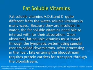
Fat Soluble Vitamins. Fat soluble vitamins include: A and carotenoids, E, K, D Associated with fat absorption Needed in small amounts Stored in fatty tissues Excess intake has toxic consequences. Retinol and caroteinoids Lipid-soluble red, orange, and yellow pigments produced by plants

E N D
Fat Soluble Vitamins • Fat soluble vitamins include: A and carotenoids, E, K, D • Associated with fat absorption • Needed in small amounts • Stored in fatty tissues • Excess intake has toxic consequences
Retinol and caroteinoids • Lipid-soluble red, orange, and yellow pigments produced by plants • Fewer than 10% have vitamin A activity • B carotene, a carotene, b cryptoxanthin • Others also have physiological importance • Lycopene • Canthaxanthin • Zeaxanthin Fig. 10-1a, p. 327
70-90% vitamin A absorption if fat is present <5% to 60% for carotenoids; vitamin E interferes CRBPII = cellular retinol binding protein LRAT = lecithin retinol acyl transferase Specfic protein carrier- vitamin A Passive diffusion - carotenoids Fig. 10-2, p. 329
‘‘Outer limiting membrane’’ Photoreceptor (rod) cell Müller cell Outer segment Inner segment Capillary Outer of photoreceptor (rod) cell Segment Pigment epithelium Nucleus • Functions: Vitamin A • Vision • Cell differentiation, growth, reproduction • Bone development • Immune system Fig. 10-7, p. 334
Functions: Vitamin A • Vision • Cell differentiation, growth, reproduction • Bone development • Immune system Fig. 10-9, p. 335
Functions: Vitamin A • Vision • Cell differentiation, growth, reproduction • Bone development • Immune system
Functions: Vitamin A • Vision • Cell differentiation, growth, reproduction • Bone development • Immune system • Function: Carotenoids • Antioxidants for singlet oxygen; • Lycopene > vitamin E > carotene > cryptoxanthin > zeaxanthin, carotene > lutein • (also work better when used together) • Antioxidant for lipid peroxides (works with vitamin E) • Lower incidence of atherosclerosis through prevention of oxidation of LDLs
Interaction with other nutrients: • Vitamins E and K (inversely related; high A, low E and K) • Zinc and iron • Protein • Excretion: most in urine as oxoretinoic acid, small amounts in expired air, some in feces Fig. 10-10, p. 339
Deficiency: • increased morbidity in children under age 5 with no evident clinical signs of deficiency • Signs, when present include xeropthalmia, anorexia, retarded growth, increased susceptibility to infections, enlargement of hair follicle, and keratinization of epithelial (mucous cells) of the skin. • Toxicity: • Hypervitaminosis A • Nausea, vomiting, double vision, headache, dizziness, and desquamation of the skin • Teratogen
Vitamin D (a seco-steroid) plants animals Fig. 10-11, p. 344
Dietary Vitamin D is absorbed from a micelle, along with other fats. • About 50% of dietary D3 is absorbed. Most absorbed in distal small intestine. • Incorporated into chylomicrons • Cholecalciferol from the skin is bound to DBP and travels primarily to the liver, but can be picked up by other tissues as well (muscle and adipose) • Blood is the major storage site; half-life of 10-21 days • Hydroxylases generate the active form of the vitamin (25-OH cholecalciferol) • Release by the kidney of active forms; a half-life of 4-6 hours in the blood Fig. 10-12, p. 346
Functions: • Acts as a steroid hormonein calcium homeostasis • Intestinal effects • Effects on the kidney • Effects on bone Fig. 10-13, p. 347
Deficiency: rickets, osteomalacia • Interaction with other nutrients: • Calcium, phosphorus, vitamin K • Excretion: • Bile > feces > urine • Toxicity: • Not possible from excess exposure to sunlight • Few cases; calcification of soft tissues, hypertension, anorexia, renal dysfunction
Only form with biologic activity Fig. 10-17, p. 353
Digestion and Transport: • Synthetic forms are de-esterified • Free alcohol forms are absorbed passively in micelles; non-saturable • 20-80% absorption; better with fats • Incorporated into chylomicrons in intestinal cell and sent out into lymph • Transfer between chylomicrons, HDLs and LDLs occurs in the blood. HDLs and LDLs contain highest concentration of the vitamin • Half-life of about 48 hrs. • Some stored in adipose, liver, lung, heart, muscle, adrenals Table 10-3, p. 354
Functions: • Maintenance of membranes - prevents oxidation of unsaturated fatty acids contained in the phospholipids (includes membranes of mitochondria and ER) • Reduced LDL oxidation; decreased plaque formation • Reduction in cataract formation • Reduced oxidation in diabetics • Suppression of activity of HMGcoA reductase (cholesterol synthesis) 1 2 Fig. 10-18, p. 356
Regeneration • Nutrient Interactions: • Function closely linked to selenium (needed for GSH peroxidase), vitamin C, sulfur containing amino acids, • Inhibits carotene absorption and conversion to retinol; may impair vitamin K absorption; may cause vitamin -D dependent bone mineralization problems • Deficiency: • Rare except in populations with fat malabsorption (cystic fibrosis) • myopathy and weakness, croid pigment accumulation, and degenerative neurologic problems • Toxicity: one of the least toxic; bleeding problems Fig. 10-19, p. 356
Vitamin K • Absorption: in micelles; incorporated into chylomicrons, then chylomicron remnants, then VLDLs, then HDLs and LDLs. • Found mainly in liver and heart. Turnover is once every 2.5 hrs. Synthetic form From green plants Synthesized by bacteria Fig. 10-20, p. 361
Functions: blood clotting and bone mineralization Fig. 10-21, p. 363
Vitamin K cycle Needed for protein carboxylation Vit. K usually only present in this form in the body Osteocalcin or Bone Gla protein Matrix Gla protein Fig. 10-23, p. 364
Deficiency: rare in adults; newborns, chronic antibiotic administration, and malabsorption can result in deficiency Bleeding episodes Osteoporosis Toxicity: none known