Muscle and movement
290 likes | 319 Views
Explore the roles of bones, ligaments, muscles, tendons, and nerves in human movement. Learn about the structure and function of key joint components and muscle fibers. Compare hip and knee movements and delve into muscle contraction mechanisms.

Muscle and movement
E N D
Presentation Transcript
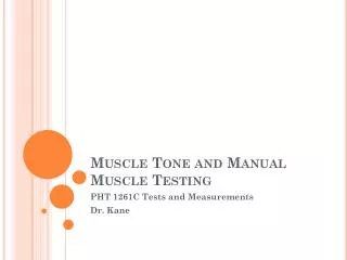
Muscle and movement Topic 11.2
Assessment statements 11.2.1 State the roles of bones, ligaments, muscles, tendons and nerves in human movement. 11.2.2 Label a diagram of the human elbow joint, including cartilage, synovial fluid, joint capsule, named bones and antagonistic muscles (biceps and triceps). 11.2.3 Outline the functions of the structures in the human elbow joint named in 11.2.2. 11.2.4 Compare the movements of the hip joint and the knee joint. 11.2.5 Describe the structure of striated muscle fibres, including the myofibrils with light and dark bands, mitochondria, the sarcoplasmic reticulum, nuclei and the sarcolemma. 11.2.6 Draw and label a diagram to show the structure of a sarcomere, including Z lines, actin filaments, myosin filaments with heads, and the resultant light and dark bands. 11.2.7 Explain how skeletal muscle contracts, including the release of calcium ions from the sarcoplasmic reticulum, the formation of cross-bridges, the sliding of actin and myosin filaments, and the use of ATP to break cross-bridges and re-set myosin heads. 11.2.8 Analyseelectron micrographs to find the state of contraction of muscle fibres.
Joints • Articulation or arthrosis, point where two or more bones contact one another • Arthrology is the scientific study of joints • Rheumatology is the branch of medicine devoted to joint disease and conditions • Kinesiology is the scientific study of the movement of the human body • Joints provide mobility and hold the body together • Include: bones, ligaments, muscles, tendons, and nerves
Bones (living organs) • Provide a hard framework to support the body • Allow protection of vulnerable softer tissue and organs • Act as levers so that body movement can occur • Forms blood cells in the bone marrow • Allows storage of minerals, especially calcium and phosphorus
Muscles and tendons • Muscles attached to bones by tendons • Tendons are cords of dense connective tissue • Arrangement of the bones and the design of the joints determine the type or range of motion possible in any particular area of the body • Muscles provide force necessary for movement by shortening the length of the fibers or cells • Occur as antagonistic pairs which allow the body part to return to its original position after movement
Ligaments and nerves • Ligaments are band-like connective tissue that serves to strengthen the joint and provide stability • Have many different types of sensory nerve endings which that help to prevent over-extension of the joint and its parts • Proprioceptors in ligaments and muscles allow constant monitoring of the position of the joint parts
Elbow joint Synovial fluid is located within the synovial cavity. This cavity is located within the joint capsule. The joint capsule is composed of dense connective tissue that is continuous with the membrane of the involved bones. Joint capsule Ends of bones lined with cartilage Synovial cavity containing synovial fluid
Types of joints • Synovial – contain synovial cavity • Diarthrotic – freely movable • Hinge – provides an opening-and-closing type of movement • Ball-and-socket – permits movement in several directions
Definitions • Flexion – decrease in angle between connecting bones • Extension – increase in angle between connecting bones • Abduction – movement of bone away from body midline • Adduction – movement of bone toward midline • Circumduction – distal or far end of a limb moves in a circle • Rotation – a bone revolves around its own longitudinal axis
Muscle • Three types: • Skeletal or striated • Cardiac • Smooth or non-striated
Striated muscle cells • Composed of thousands of cells, which are called muscle fibers b/c of their elongated shape • Blood vessels and nerves penetrate the muscle body • Each muscle fiber contains multiple nuclei that lie just inside the plasma membrane, which is called the sarcolema
Sarcolemma has multiple tunnel-like extensions that penetrate the interior of the cell called transverse or T tubules • cytoplasm of muscle fibers is called the sarcoplasm • Sarcoplasm contains large numbers of glycosomes that store glycogen • Sarcoplasm also contains large amounts of myoglobin
Sarcoplasmic reticulum is a fluid-filled system of membranous sacs surrounding the muscle myofibrils • Myofibrils are rod-shaped bodies that run the length of the cell and are the contractile elements of the muscle cell • Myofibrils run parallel to one another and have numerous mitochondria squeezed between them
Myofibrils • Made up of sarcomeres which allow movement • Often described as banded: • Z lines mark the ends of the sarcomere • A bands are dark in color and extend the entire length of the myosin filaments; narrow H band occurs in the middle containing only myosin, no actin; supporting protein occurs in the middle of myosin producing M line • I bands are light in color and contain only actin, no myosin
Two types of filaments or myofilaments that cause the banded appearance of the muscle fiber • These two myofilaments are composed of two contractile proteins, actin and myosin
Muscle Contraction • Explained by the sliding filament theory proposed by Hugh Huxley in 1954 • States that muscles contract when actin myofilaments slide over myosin myofilaments
Sliding Filament Theory • Motor neuron carries an action potential until it reaches a neuromuscular junction • Neurotransmitter called acetylcholine is released into the gap between the axon terminal and the sarcolemma of the muscle fiber • Acetylcholine bines to receptors in the sarcolemma • Sarcolemma ion channels open and sodium ions move through the membrane
Muscle action potential is generated • Muscle action potential moves along the membrane and through the T tubules • Acetylcholine is broken down by acetylcholinesterase • Muscle action potential moving along T tubule causes release of calcium ions from the sarcoplasmic reticulum. Calcium ions flood into the sarcoplasm
Calcium ions bind to troponin on the actinmyofilaments. This exposes the myosin-binding sites • Myosin heads include ATPase which splits ATP and releases energy • Myosin heads then bind to the myosin-binding sites on the actin with the help of the protein called tropomyosin • Myosin-actin cross-bridges rotate toward the center of the sarcomere. This produces the power or working stroke.
ATP once again binds to the myosin head resulting in the detachment of myosin from the actin • If there are no further action potentials, the level of calcium ions in the sarcoplasm falls. The troponin-tropomyosin complex then moves to its original position, thus blocking the myosin-binding sites. The muscle then relaxes.
Useful websites • http://3dotstudio.com/zz.html • http://www.blackwellpublishing.com/Matthews/myosin.html • http://entochem.tamu.edu/musclestruccontractswf/index.html • http://highered.mcgraw-hill.com/sites/0072495855/student_view0/chapter10/animation__sarcomere_contraction.html
Rigor mortis • After death, calcium ions leak out of the sarcoplasmic reticulum and bind to troponin • This allows actin to slide, but lack of ATP production prevents myosin heads from detaching from the actin • Result is rigidity for about 24 hours unter further muscle deterioration occurs
When the muscle is maximally contracted, the H zone disappears, the Z lines move closer together, the I bands are no longer present, and the A bands appear to run the complete length of the sarcomeres • Can be in various states of partial contraction • This causes a difference in the position of the sarcomere parts • The number of muscle fibers in a muscle going through contraction determines the overall strength of a muscle contraction