Muscle: enables movement possible
500 likes | 931 Views
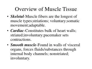
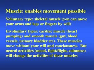
Muscle: enables movement possible Voluntary type: skeletal muscle (you can move your arms and legs or fingers by will)

Muscle: enables movement possible
E N D
Presentation Transcript
Muscle: enables movement possible Voluntary type: skeletal muscle (you can move your arms and legs or fingers by will) Involuntary types: cardiac muscle (heart pumping) and smooth muscle (gut, blood vessels, urinary bladder etc). These muscles move without your will and conciousness. But neural activities (mood, fight/flight, calmness) will change the activities of these muscles
Striations or not Striated Non-striated Smooth muscle Skeletal muscle Cardiac muscle
The sliding filament mechanism, in which myosin filaments bind to and move actin filaments, is the basis for shortening of stimulated skeletal, smooth, and cardiac muscles. • In all three types of muscle, myosin and actin interactions are regulated by the availability of calcium ions. • Changes in the membrane potential of muscles are linked to internal changes in calcium release (and contraction).
Skeletal muscle • A single skeletal muscle cell is called a muscle fiber • Each muscle fiber is formed during development by the fusion of a number of undifferentiated, mononucleated cells, known as myoblasts, into a single cylindrical multinucleated cell. • The term muscle = a number of muscle fibers bound together by connective tissue. Muscles are attached to bones by tendons
The sarcomere is composed of: thick filaments called myosin, anchored in place by titin fibers, and thin filaments called actin, anchored to Z-lines .
A cross section through a sarcomere shows that: • each myosin can interact with 6 actin filaments, and • each actin can interact with 3 myosin filaments.
Contraction refers to force generation in the muscle fibers, not necessarily means shortening. • Example, holding a heavy book with your arms straight requires muscle contraction but not shortening (isometric contraction) • But if you move the heavy book upward, it involves shortening (isotonic contraction)
Contraction involving shortening: myosin binds to actin, and slides it, pulling the Z-lines closer together, and reducing the width of the I-bands. Note that filament lengths have not changed.
Contraction: myosin’s cross-bridges bind to actin; the crossbridges then flex to slide actin.
The thick filament called myosin is actually a polymer of myosin molecules, each of which has a flexible cross-bridge that binds ATP and actin.
If Ca present, cycle repeats. 3,4 are distinct steps!
In relaxed skeletal muscle, tropomyosin blocks the cross-bridge binding site on actin. Contraction occurs when calcium ions bind to troponin; this complex then pulls tropomyosin away from the cross-bridge binding site.
The latent period between excitation and development of tension in a skeletal muscle includes the time needed to release Ca++ from sarcoplasmic reticulum, move tropomyosin, and cycle the cross-bridges.
The transverse tubules bring action potentials into the interior of the skeletal muscle fibers, so that the wave of depolarization passes close to the sarcoplasmic reticulum, stimulating the release of calcium ions.
The extensive meshwork of sarcoplasmic reticulum assures that when it releases calcium ions they can readily diffuse to all of the troponin sites.
Passage of an action potential along the transverse tubule. Note that TT is very close to SR. Depolarization is sensed by DHP receptor, which opens the coupled voltage-gated calcium channels, the “ryanodine receptor,” located on the sarcoplasmic reticulum, and calcium ions released into the cytosol bind to troponin. Note that after excitation, Ca is pumped back to SR.
Nerve cells that innervte skeletal muscle fibers are called motor neurons. A single motor unit consists of a motor neuron and all of the muscle fibers it controls.
The neuromuscular junction is the point of synaptic contact between the axon terminal of a motor neuron and the muscle fiber it controls. Action potentials in the motor neuron cause acetylcholine release into the neuromuscular junction. Muscle contraction follows the delivery of acetylcholine to the muscle fiber.
Mechanical response of a single muscle fiber to a single action potential is called a twitch. iso = same tonic = tension metric = length In isotonic contraction, latent period is considerably longer, as shortening will not occur until muscle tension > load * Before shortening is isometric contraction Heavier load, longer latent period Contraction is isometric when load is too heavy *
All three are isotonic contractions. Light loads are more rapidly moved than heavy loads.
T e m p o r a l s u m m a t i o n. Complete dissipation of elastic tension between subsequent stimuli. S3 occurred prior to the complete dissipation of elastic tension from S2. S3 occurred prior tothe dissipation of ANY elastic tension from S2.
Unfused tetanus: partial dissipation of elastic tension between subsequent stimuli. Fused tetanus: no time for dissipation of elastic tension between rapidly recurring stimuli. Here continuous stimulation constantly releases Ca so Ca is not effectively pumped back to SR, resulting in prolonged Ca elevation
Short sarcomere: actin filaments lack room to slide, so little tension can be developed. Optimal-length sarcomere: lots of actin-myosin overlap and plenty of room to slide. Relaxed skeletal muscle fibers are near lo Long sarcomere: actin and myosin do not overlap much, so little tension can be developed.
At rest, muscle fibers build up creatine phosphate as sources of ATP: initial reserve for a few seconds. Later on resort to glycolysis and oxidative phosphorylation. When muscular activities is prolonged and intense, anaerobic glycolysis begins to provide major source of ATP with the generation of lactic acid. Also, oxygen debt has to be repaid to metabolize lactic acid and to regenerate creatine phosphate and ATP.
Rest overcomes fatigue, but fatigue will reoccur sooner if inadequate recovery time passes. In skeletal muscle, repetitive stimulation leads to fatigue, evident as reduced tension. 3 mechanisms may account for the fatigue: Accumulationof K ions in the T tubules during repolarization Lactic acid (acidity) inhibits contractile machineries eg. Actin/myosin, Ca release channels ADP and Pi inhibits cross-bridge cycling.
3 major types of skeletal muscles Slow-oxidative skeletal muscle responds well to repetitive stimulation without becoming fatigued; muscles of body posture are examples. Low myosin ATPase activities and high oxidative capacity. Fast-oxidative skeletal muscle responds quickly and to repetitive stimulation without becoming fatigued; muscles used in walking are examples. High myosin ATPase activities and high oxidative capacity. Fast-glycolytic skeletal muscle is used for quick bursts of strong activation, such as muscles used to jump or to run a short sprint. High myosin ATPase activities and high glycolytic capacity. Most skeletal muscles include allthreetypes.
Fast glycolytic fibers are larger in diameter and are therefore more forceful. However, easily fatigued. Good for “explosive”sports like high jumping or sprint.
Allthreetypesof muscle fibers are represented in a typical skeletal muscle, and, under tetanic stimulation, make the predicted contributions to the development of muscle tension. Fast- glycolytic Fast-oxidative In increasing muscle strength, Recruitment of small motor neurons first (they activate slow-oxid. Fibers) and finally large motor neurons (they activate fast glycolytic fibers). Slow-oxidative
For delicate movements (eye and hand muscles), about 13 fibers innervated by a motor neuron. For leg muscles, hundreds to thousands of fibers in a motor unit.
Exercise or NOT • Denervation atrophy: when the innervating neuron or neuromuscular junctions is destroyed or non-functional, muscle receive no stimulation and will become smaller in diameter and will have less contractile proteins. • Disuse atrophy: atrophy happens when a muscle is not used for a long time. • Exercise causes hypertrophy and enhanced ATP production capacity. • Remember, in atrophy or hypertrophy, no change in muscle fiber number, but in SIZE, and metobolic capacity.
Clincal cases of muscle abnormalities • Muscle cramps: involuntary tetanic contraction. Overexercise may cause electrolyte imbalance in the extracellular fluid surrounding muscle and nerve. These cause abnormally high neuron firing rate. • Hypocalcemic tetany: involuntary tetanic contraction as extracellular Ca concentration drops to 40% of normal value. This condition increases membrane excitability (Na influx)
Clincal cases of muscle abnormalities • Muscular dystrophy: a protein called dystrophin is missing. It is similar to cytoskeletal protein, maintaining plasma membrane and cell structural integrity. Progressive degeneration of skeletal and cardiac muscles, eventually early death before 20. • Myasthenia gravis: muscle fatigue and weakness due to destruction of nicotinic Ach receptor of motor end plate. Reason: auto-antibodies to the receptors. Reduced end-plate potential.
Duchenne muscular dystrophy weakens the hip and trunk muscles, thus altering the lever-system relationships of the muscles and bones that are used to stand up.
Smooth muscle • Innervated by automomic nervous system, therefore involuntary • Actin-myosin contractile machineries, but organisation and excitation-contraction coupling is different from skeletal muscle.
Thick (myosin-based) and thin (actin-based) filaments, biochemically similar to those in skeletal muscle fibers, interact to cause smooth muscle contraction. Troponin is ABSENT in SMC.
Ca binds to calmodulin (CM) and then activates myosin light chain kinase (MLCK) MLCK phosphorylates globular head of MLC. The latter binds to actin and slides. Myosin ATPase has a very low rate, therefore contraction velocity is much slower than skeletal muscle. Note another ATP molecule is to detach myosin from actin, and its hydrolysis re-energizes the myosin head.
1 2 • Sources of calcium very different: • Opening of voltage-dependent Ca channel • RACC • Second messenger-triggered Ca release 3
Some SMC are pacemaker cells Rhythmic changes in the membrane potential of smooth muscles results in rhythmic patterns of action potentials and therefore rhythmic contraction; in the gut, neighboring cells use gap junctions to further coordinate these rhythmic contractions.
Neuronal and hormonal influences • Some neurotransmitters increase, while some other decrease, SMC contractility • The same neurotransmitter can produce opposite effects in different types of SMC: eg. Noradrenaline contracts most SMC by acting on alpha- adrenergic receptors, but relaxes airway SMC by acting on beta-2 adrenergic receptors. • Hormones, in a similar manner, may either stimulate or inhibit SMC contraction.
Other factors • Local factors will also affect SMC contractility to tell the cell’s immediate environment. Nitric oxide released from endothelial cells relaxes SMC. • Stretch opens mechanosensitive ion channels which then cause membrane depolarization.