Isolation of Nucleic Acids
340 likes | 957 Views
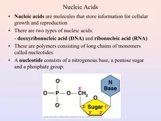
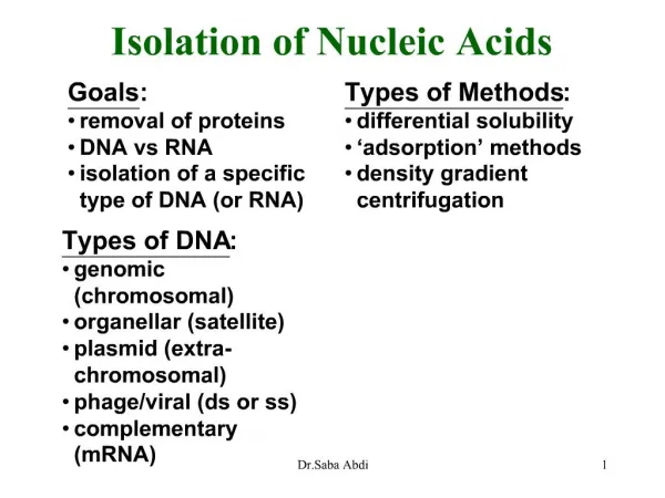
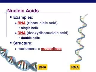
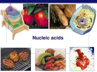
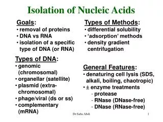
Isolation of Nucleic Acids. Goals : removal of proteins DNA vs RNA isolation of a specific type of DNA (or RNA). Types of Methods : differential solubility ‘adsorption’ methods density gradient centrifugation. Types of DNA : genomic (chromosomal) organellar (satellite)

Isolation of Nucleic Acids
E N D
Presentation Transcript
Isolation of Nucleic Acids • Goals: • removal of proteins • DNA vs RNA • isolation of a specific type of DNA (or RNA) • Types of Methods: • differential solubility • ‘adsorption’ methods • density gradient centrifugation • Types of DNA: • genomic (chromosomal) • organellar (satellite) • plasmid (extra-chromosomal) • phage/viral (ds or ss) • complementary (mRNA) • General Features: • denaturing cell lysis (SDS, alkali, boiling, chaotropic) • enzyme treatments • protease • RNase (DNase-free) • DNase (RNase-free) Dr.Saba Abdi
aqueous phase (nucleic acids) phenol phase (proteins) High MW Genomic DNA Isolation • Typical Procedure • Cell Lysis • 0.5% SDS + proteinase K (55o several hours) • Phenol Extraction • gentle rocking several hours • Ethanol Precipitation • RNAse followed by proteinase K • Repeat phenol extrac-tion and EtOH ppt • Phenol Extraction • mix sample with equal volume of sat. phenol soln • retain aqueous phase • optional chloroform/isoamyl alcohol extraction(s) Dr.Saba Abdi
High MW Genomic DNA Isolation • Typical Procedure • Cell Lysis • 0.5% SDS + proteinase K (55o several hours) • Phenol Extraction • gentle rocking several hours • Ethanol Precipitation • RNAse followed by proteinase K • Repeat Phenol Extrac-tion and EtOH ppt • EtOH Precipitation • 2-2.5 volumes EtOH, -20o • high salt, pH 5-5.5 • centrifuge or ‘spool’ out Dr.Saba Abdi
Isolation of RNA Special Considerations • RNAse inhibitors! • extraction in guanidine salts • phenol extractions at pH 5-6 • (pH 8 for DNA) • treatment with RNase-free DNase • selective precipitation of high MW forms (rRNA, mRNA) with LiCl • oligo-dT column Dr.Saba Abdi
Adsorption Methods • nucleic acids selectively absorb to silica or resins in the presence of certain chaotropic agents or salts Plasmid Miniprep Protocol 1. Solubilize bacteria in alkali solution 2. Neutralize with Na-acetate 3. Centrifuge, discard pellet 4. Mix supernatant with resin + chaotropic agent 5. Wash resin 6. Elute DNA with low salt buffer • applications: • plasmid preps • fragments after electrophoresis • PCR templates Dr.Saba Abdi
1.74 1.72 density (g/cm3) 1.70 1.68 20 60 80 40 % GC base pairs Density Gradient Centrifugation • rate zonal/sucrose (size fractionation) • electrophoresis more common • isopycnic/CsCl (density) • DNA ~1.7 g/cm3 • protein ~1.3 g/cm3 • RNA > DNA • ssDNA > dsDNA • GC content Dr.Saba Abdi
CsCl Gradients • Applications • large scale preparations • high purity • ‘satellite’ DNA • RNA ‘cushions’ CsCl Gradients Dr.Saba Abdi
Using Spectroscopy to analyze DNA DNA absorbs UV light with a major peak at 260 nm This absorption is useful because it varies with the structure of DNA (&RNA) i.e. extinction coefficient depends on the structure Optical Density Wave Length 260 dsDNA Low extinction coefficient ssDNA Higher extinction coefficient Dr.Saba Abdi
Evaluation of Nucleic Acids • spectrophotometrically • quantity • quality • fluorescent dyes • gel electrophoresis Dr.Saba Abdi
Agarose Gel Stained with ethidium bromide (EtBR) to Visualize the DNA slots where DNA is loaded 1000 bp 700 bp 600 bp 500 bp Screening PCR products to test for the presence of specific DNA sequences molecular weight markers correct PCR product molecular weight markers Dr.Saba Abdi
Intercalating Agents Distort the Double Helix Several hydrophobic molecules containing flat aromatic and fused heterocyclic rings can insert between the stacked base pairs of DNA. These molecules are called intercalating agents. Intercalating agents are potential Cancer-inducing reagents. Dr.Saba Abdi
Two Methods: • chemical cleavage xxx (Maxam and Gilbert) • synthetic oligonucleotides • GC-rich DNA • dideoxy (Sanger) • based on 2’3’-dideoxynucleotides as chain terminators ì H DNA Sequencing Dr.Saba Abdi
Dideoxy Chain Termination Dr.Saba Abdi
DNA sequencing: the Sanger (dideoxy) method Figure 7-29b,c Dr.Saba Abdi
NTP, dNTPs and ddNTPs Dr.Saba Abdi
DNA sequencing: the Sanger method Four separate polymerization reactions are performed Figure 7-29a Dr.Saba Abdi
DNA Sequencing Dr.Saba Abdi
Reading a DNA Sequencing Gel Sequence 5’ to 3’ C G G G C G T Dr.Saba Abdi
Semi-Automated Sequencing • thermal cycler • fluorescent ddNTPs • unique spectra • measure intensity of DNA products on gel è Dr.Saba Abdi
Automated DNA Sequencing with Fluorescent Dyes Each different ddNTP is coupled to a different colored fluorescent dye ddTTP is red; ddGTP is black etc. Dr.Saba Abdi