Download
1 / 20
270 likes | 674 Views
A Technical Seminar On . X-RAY AND CT SCAN. Introduction. X-rays were discovered by the German physicist Will elm Konrad Rontgen in November 1895. He called the 'new kind of ray' or X-rays( X for the unknown).their usefulness to visualize the internal anatomy of humans was established.

E N D
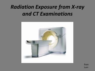
A Technical Seminar On X-RAY AND CT SCAN
Introduction • X-rays were discovered by the German physicist Will elm Konrad Rontgen in November 1895. He called the 'new kind of ray' or X-rays( X for the unknown).their usefulness to visualize the internal anatomy of humans was established. • Today, imaging with X-rays is perhaps the most commonly used • diagnostic tool with the medical profession, and the techniques from a • simple chest radiography to a digital subtraction angiography or computertomography depend on the use of X-rays.
What is X-ray? • X-radiation (composed of X-rays) is a form of electromagnetic radiation. • X-rays have a wavelength in the range of 0.01 to 10 nanometers. • X-ray photons carry enough energy to ionize atoms and disrupt molecular bonds.
Types • X-rays is of two types • Hard X-rays • X-rays with photon energies above 5-10 keV (below 0.2-0.1 nm wavelength). • Soft X-rays • X-rays with photon energies below 5-10 keV (below 0.2-0.1 nm wavelength).
Properties of x-ray • X-ray photons carry enough energy to ionize atoms and disrupt molecular bonds. The ionizing capability of X-rays can be utilized in cancertreatment to kill maligant cells using radiation therapy. • Ability for producing luminescence. • Effect in case of photographic emulsions. • Capability for penetration of matter in various materials.
Medical uses • Computer tomography • Imaging alternatives for soft tissues are computed axial tomography (CAT or CT scanning) • Fluoroscopy • Fluroscopy is another X-ray test methodology. Examples include cardiac catheterization (to examine for coronary artery blockages) and Barium swallow (to examine for esophageal disorders). • Radiotherapy • The use of X-rays as a treatment is known as radiation therapy and is largely used for the management (including palliation of cancer); it requires higher radiation energies than for imaging alone.
Principle of Working X-rays are produced in: specially constructed glass tube, which basically comprises. • A source for the production 0f electrons, • a energy source to accelerate the electrons, • a free electron path, • a means of focusing the electron beam and • a device to stop the electrons. Stationary mode tubes and rotating anode tubes are the two main types of X-ray tubes:
Failure with X-ray • There are two main limitations of using conventional X-rays to examine internal structures of the body. • Firstly, the super-imposition of the three-dimensional information onto a single plane makes diagnosis confusing and often difficult. • Secondly, the photographic film usually used for making radiographs has a limited dynamic range and, therefore, only objects that have large variations inX-ray absorption relative to their surroundings will cause sufficient contrast differences on the film to be distinguished by the eye.
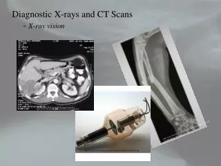
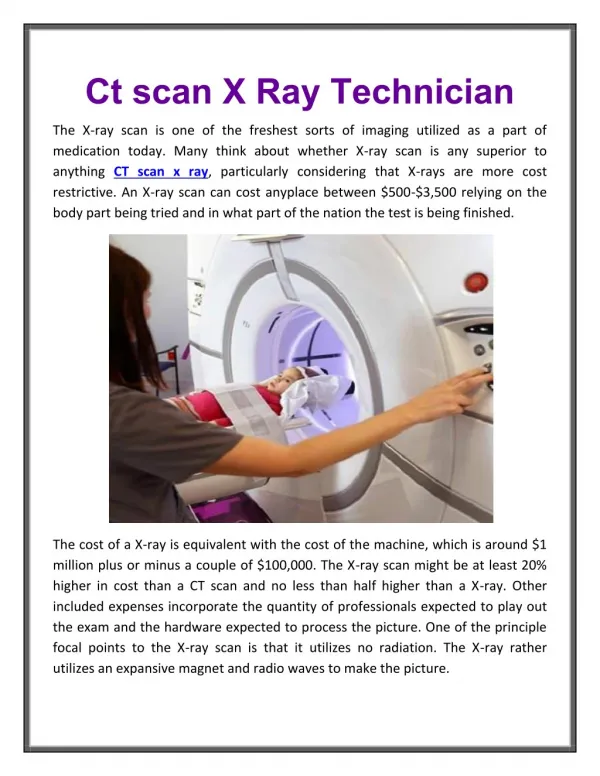
What is CT-Scan? • A computed axial tomography scan or CT Scan (also CAT Scan) is a medical imaging technology that uses finely focused X-rays together with a detector rather than photographic film combined with computer imaging technology to create detailed three-dimensional images of interior body tissue, including soft tissue. It is primarily used to detect and locate structures within the body that cannot be located by other forms of radiological investigation.
Basic Principles of CT scan • Mathematical principles of CT were first developed in 1917 by Radon • Proved that an image of an unknown object could be produced if one had an infinite number of projections through the object • Plain film imaging reduces the 3D patient anatomy to a 2D projection image
Density at a given point on an image represents the x-ray attenuation properties within the patient along a line between the x-ray focal spot and the point on the detector corresponding to the point on the image • With a conventional radiograph, information with respect to the dimension parallel to the x-ray beam is lost • Limitation can be overcome, to some degree, by acquiring two images at an angle of 90 degrees to one another • For objects that can be identified in both images, the two films provide location information
Working of CT SCAN • In principle, computed tomography involves the determination of attenuation characteristics for each small volume of tissue in the patient slice, which constitute the transmitted radiation intensity recorded from various irradiation directions. It is these calculated tissue attenuation characteristics that actually compose the CT image. • For a monochromatic X-ray beam, the tissue attenuation characteristics can be described by • It = Io e- x • I0 = Incident radiation intensity • It = Transmitted intensity • x = Thickness of tissue • = Characteristic attenuation coefficient of tissue
System Components All computed tomography systems consist of the following four major sub-systems: • Scanning system—This takes suitable readings for a picture to be reconstructed, and includes X-ray source and detectors. • Processing unit—This converts these readings into intelligible picture information. • Viewing part—It presents this information in visual form and includes other manipulative aids to assist diagnosis. • Storage unit—This enables the information to be stored for subsequent analysis.
Advntages of CT-scan over X-ray • Traditional X-rays suffer from their inability to give any perception of depth to the physician. This is because the X-ray can only picture the average density of any tissue in front of the X-ray film. A CT Scan gets around this limitation in three ways: • - Instead of using a broad beam of X-rays, a CT Scan uses a very narrowly focused beam of X-rays that can only penetrate straight through the body in a straight line to the detector. • - The X-ray source is rotated around the body so that the X-rays pass through the entire structure in both directions. • - A computer is used to reconstruct the intensity of the X-rays into an image showing the density of any point of the plane through which the X-rays passed.