Download
1 / 14
160 likes | 292 Views
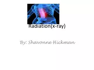
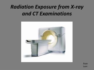
Radiation Exposure from X-ray and CT Examinations. Evan Lum. What is an X-ray. An x-ray is a form of electromagnetic radiation which falls in between gamma rays and ultra violet rays on the electromagnetic spectrum. They have a wavelength of anywhere between 0.01 nm and 10 nm.

E N D
What is an X-ray • An x-ray is a form of electromagnetic radiation which falls in between gamma rays and ultra violet rays on the electromagnetic spectrum. • They have a wavelength of anywhere between 0.01 nm and 10 nm. • Corresponds to an energy of 0.1 to about 123 keV (kiloelectron volts)
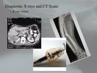
What is a CT scan? • CT stands for computed tomography which is a medical imaging method employing tomography (imaging in sections) created by computer processing. • Using a series of x-ray images about a single axis of rotation, CT produces volume data such as 3D images of internal organs which can be manipulated through a process called “windowing”.
Medical Uses for X-ray • Plain x-rays are mostly used to detect pathology in the skeletal system. • Sometimes used for soft tissue: • Chest x-ray to identify lung diseases like pneumonia or lung cancer. • Abdominal x-ray to detect intestinal obstruction, free air or free fluid.
Medical Uses for CT Scan • Head – detect infarction, tumors, hemorrhage, bone trauma. • Lungs – shows internal aspects of the lungs which plain x-ray can not. • Cardiac – excellent imaging of coronary arteries • Abdominal/Pelvic – sensitive method for diagnoses of abdominal diseases. • Extremities – can image complex fractures, especially around joints. Also ligament injuries and dislocations can be seen.
Radiation Exposure • The Sievert is the SI unit for radiation dose which is equivalent to one joule per kilogram. • The average American is exposed to about 3 mSv per year from naturally occurring radioactive materials and cosmic radiation from outer space. • This amount of radiation exposure over such a long period of time is basically harmless to us.
Radiation from X-rays • Extremities – 0.001 mSv per x-ray (not harmful) • Spine – 1.5 mSv per x-ray (about 6 months worth of natural exposure) • GI tract – 6-8 mSv per x-ray (2-3 years worth of natural exposure) • Each of these are considered not to be relatively harmful
Radiation from CT Scan • Head – 2 mSv per CT scan (under 1 year) • Spine – 6 mSv per CT scan (2 years) • Chest – 7 mSv per CT scan (over 2 years) • Abdomen/Pelvis – 15 mSv per CT scan (5 years) • Abdomen/Pelvis without contrast material – 30 mSv per CT scan (10 years) • Coronary Computed Tomography Angiography (CTA) – 16 mSv (5 years)
Why is this dangerous? • Although radiation can be used to treat cancer, it can also cause it. • The delivery of energy waves through our body can alter atoms’ molecular structure causing cells in our body to mutate and become cancerous.
Statistics • Higher risk procedures can leave patients with up to a 1 in 500 chance of getting cancer. • The united states gives over 60 million CT scans per year. • It has been estimated recently that CT scanning has been the cause of about 30,000 cases of cancer yearly.
What Can We Do? • Due to their major contribution to the medical field, the benefits of x-ray imaging and CT scanning usually outweigh the risks from radiation exposure. • Something we can do to avoid this is avoiding unnecessary procedures. • This decision should only be made by a professional not a patient!
Sources • http://www.usatoday.com/news/health/2009-12-15-radiation15_st_N.htm • http://en.wikipedia.org/wiki/X-ray_computed_tomography • http://www.radiologyinfo.org/en/pdf/sfty_xray.pdf • http://www.worldculturepictorial.com/blog/content/ct-scan-study-shows-increased-radiation-exposure-cancer-risks-tests-often-unnecessary • http://www.hanskellner.com/2005/06/27/left-shoulder-xray/ • “The Physics of Medical Imaging”, by S. Webb, New York: Adam Hilger, 1990.