Download
1 / 89
890 likes | 1.14k Views
Neurological System Chapter 38. White Christensen Kockrow Adam Leslie Lehmkuhl, RN 2008. Affect Agnosia AneurysmAphasia Areflexia Apraxia Ataxia Aura Automatism Autonomic nervous system. Bradykinesia Stroke Central nervous system Diplopia. Key Terms. Cephalgia

E N D
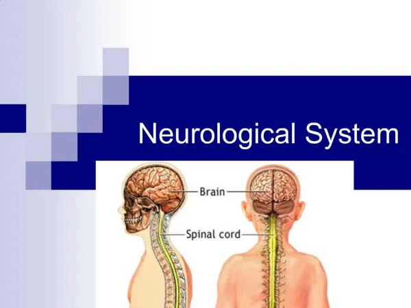
Neurological SystemChapter 38 White Christensen Kockrow Adam Leslie Lehmkuhl, RN 2008
Affect Agnosia AneurysmAphasia Areflexia Apraxia Ataxia Aura Automatism Autonomic nervous system Bradykinesia Stroke Central nervous system Diplopia Key Terms
Cephalgia Chorea Copralalia Dysarthria Dysphagia Emotional lability Encephalitis Decorticate posturing: of arms and legs rigid plantar flexion Decerebrate: rigid extension of arms and legs with wrists turned outward Terms
Fasciculation Flaccid Glascow coma scale Global cognitive dysfunction Graphesthesia Hemanopia Hemiplegia Hemiparesis Hyperreflexia Meningitis Mentation Nystagmus Neuralgia Neurogenic shock Orientation Paraplegia Terms
Postictal period Proprioception Quadrspelegia Sclerotic Spastic Spinal shock Status epilepticus Sterognosis Unilateral neglect Vertigo Terms
The nervous system is the body’s communication network. It coordinates and organizes the functions of all other body systems. A highly complex and coordinates and controls all motor, sensory and autonomic functions. This intricate network has 2 main divisions: Central Nervous System Peripheral Nervous System Introduction
Central nervous system (CNS) Brain and spinal cord (body’s control center) Peripheral nervous system (PNS) Contains cranial and spinal nerves that connect to CNS to remote body parts which relay and transmit messages Somatic nervous system Sends messages from the CNS to the skeletal muscles; voluntary Autonomic nervous system Sends messages from the CNS to the smooth muscle, cardiac muscle and certain glands; involuntary. Includes the sympathetic and parasympathetic *note: a & i are vowels Nervous System Division
Brain is composed of gray and white matter. Gray matter is the outside which contains billions of neurons. The white matter make up the inner structure of the brain contains pathways that transmit nerve impulses to different areas of the brain. The brain and spinal cord are protected by the bony skull and vertebre, CSF and three membranes: Dura mater Pia mater Arachnoid mater CNS
Dura mater – a tough fibrous, leatherlike tissue composed of two layers. Inner lining/ layer of the skull Thick layer which covers the brain and provides support and protection Pia mater- connective tissue that covers and contours the spinal tissue and brain. Arachnoid mater- thin, fibrous membrane that hugs the brain and spinal cord, though not as preciously as the pia mater. Dura mater,Pia mater, Arachnoid mater
Between the dura mater and arachnoid membrane is the subdural space Between the pia matter and the arachnoid membrane is the subarachnoid space. Within the subarachnoid space and the brain’s four ventricles is CSF, a liquid composed of water, and traces of organic material (protein, glucose, and minerals. The fluid protects the brain and special tissue from jolts and blows The Spaces Between
Cells of the nervous system Neuron (fundamental unit of the nervous system) Delicate threadlike nerve fibers called axons and dendrites that extend from the cell body CNS
Neurons (Nerve Cell) Consist of three main parts—dendrites; cell body of neuron; and axon Dendrites conduct impulses to cell body of neuron Axons conduct impulses away from cell body of neuron Most neurons have multiple dendrites but only one axon. Gap between each neuron is a synapse and neurotransmitters conduct impulses through the gap Neurotransmitters: acetycholine, norepinephrine, dopamine, serotonin CELLS OF THE NERVOUS SYSTEM
The cerebrum is divided into right and left hemispheres The right side controls the left side of the body The left side controls the right side of the body Brain Hemispheres “Right Controls the Left, and the Left Controls the Right”
Right Hemisphere- perception, physical environment, art, music, spiritual, non-verbal communication Left Hemisphere- Analysis, interpretation, calculation, problem solving, writing, and reading Brain Hemispheres
17 to 18inches long is a 2 way conductor pathway between the brain and peripheral nervous system. Spinal cord conducts impulses to and from the brain, serves as a center for reflex action 31 pairs of spinal nerves originate from the spinal cord to the body The spinal cord has an H shaped appearance called horns. (gray mater) These horns contain the cell bodies of neurons needed for voluntary reflex action Spinal Cord
500 milliliters are produced daily CSF absorbs shock and bathes the brain The nutrients (protein, glucose, Na+urea) are delivered to the CNS cells Toxic and waste products are removed. CSF
Nerve—bundle of peripheral axons Tract—bundle of central axons White matter—tissue composed primarily of myelinated axons (nerves or tracts). Transmits nerve impulses to different areas of the brain. Gray matter—tissue composed primarily of cell bodies and unmyelinated fibers Nerve coverings—fibrous connective tissue Endoneurium—surrounds individual fibers within a nerve Perineurium—surrounds a group (fascicle) of nerve fibers Epineurium—surrounds the entire nervea NERVES AND TRACTS
Divisions of the brain Brainstem Consists of three parts of brain; named in ascending order: the medulla oblongata, pons, and midbrain Structure—white matter with bits of gray matter scattered through it Function—gray matter in the brainstem functions as reflex centers (e.g., for heartbeat, respirations, and blood vessel diameter) Sensory tracts in the brainstem conduct impulses to the higher parts of the brain Motor tracts conduct from the higher parts of the brain to the spinal cord CENTRAL NERVOUS SYSTEM
Somatic Nervous System Connects CNS to skin and skeletal muscles Conscious activities (walking, exercise) Autonomic Nervous System Connects CNS to visceral organs (e.g. heart, stomach, GI, and other viseral organs) Unconscious activities (breathing) Peripheral Nervous System
Sympathetic System Parasympathetic System ANS See pg 1064 Table 38-2
Sympathetic nervous system increases heart rate, blood pressure, dry mouth Fight or flight system Parasympathetic slows the system for normal function Decreases heart rate, decreases blood pressure.. ANS
NEUROLOGICAL ASSESSMENT • History • Cerebral function • Cranial nerve function • Motor function • Sensory function • Reflexes
History of incident or accident headaches, changes of vision, seizure activity,numbness or tingling in an extremity, mood changes, personality changes, fatigue, pupil size and reaction, level of consciousness, perception, speech, lethargy, motor coordination, proprioception Nursing assessment
Level of consciousness (responsiveness and orientation), most important indicator of change in LOC.. Call pt by name If no response… touch pt gently or shaking shoulder If no response…. use strong stimulation (e.g. nail bed pressure) To document use Glasgow Coma Scale or document pts state of arousal Alert Disorientation Lethargic Obtunded Stuporous Semiconcious Comatose Cerebral Function Assessment
Scale of responses to eye opening, motor response and verbal response with a number for each Eyes open: 4- spontaneous 3- to speech 2- to pain 1- none Best verbal response: 5- obeys commands 4- confused 3- inappropriate 2- incomprehensible 1- none Best motor response: 5- obeys commands 4- localizes pain 3- flexion to pain 2- extension to pain 1- none Total 3 to 15 <7 is considered a comatose state It is important to monitor any downward trend in the patients score. If this happens, the nurse must act quickly, call MD and assist with measures to prevent or reduce ICP and prevent further brain damage.. Glasgow Coma Scale
Mental status (e.g., Mood, behavior, facial expressions, gestures) Intellectual function (e.g., concentration and recall) repeating numbers-recall adding small numbers -calculation last meal eaten Short term memory=repeating 3 numbers stating what was eaten for last meal Long term memory=school attended time served in the military concentration general knowledge repeating recent news information Cerebral Function Assessment
Emotional status (affect). Pupillary reaction (size and equality) Cerebral Function Assessment
PERRLA: Pupils Equal Round Reactive Brisk Sluggish Non-reactive consensual Accommodation normal findings Distance= dilation Close up= constriction Cerebral Function Assessment
Oral and written communication. Vocabulary used- Aphasia: no speech Sensory aphasia: Receptive aphasia inability to comprehend the spoken or written word Motor aphasica: Expressive aphasia inability to use words or symbols Global aphasia: inability to understand the written word or to speak Anomia: inability to name objects Dysarthria: difficult speech Cerebral Function Assessment
a reflection of brain stem activity, is usually assessed by a physician or advanced practice nurse. See “Understanding Cranial Nerves” handout provided. Cranial Nerve Function Assessment
Muscle size and symmetry Compare bilaterally Muscle tone Normal, flaccid (hypotonic), rigid (constant stae of spacicity), spastic (rigid, may have tremors) Muscle strength 0 to 5 (see next slide) Push against resistance Coordination Cerebellum functioning finger to nose (38-6) run heel of foot down opposite shin (38-7) Ataxia is the inability to perform voluntary muscle function Motor Function Assessment
Muscle Strength • 5/5 strong • 4/5 fair to moderate strength • 3/5 just able to overcome gravity • 2/5 can move but not overcome gravity • 1/5 minimal power strength • 0/5 no movement
Balance Romberg test Eyes closed Feet together Arms extended in front Slight swaying is normal use safety precautions (stand in front of pt)and prevent falls Posturing Decorticate posturing: of arms and legs rigid plantar flexion Decerebrate: rigid extension of arms and legs with wrists turned outward Flaccid weak, lack muscle tone Spastic sudden involuntary movement Motor Function Assessment
Tactile Sensation use cotton ball on arms, hands, feet, legs bilat. Pain and temperature transmit on same pathway. Use safety pin dull sharp Vibration tuning fork feel vibrations on wrists and ankles Proprioception space position in regard sto joints. Passively move pt fingers or extremities and ask direction moved to pt. Unilateral neglect individual ignores one side of body Hemianopia defect in vision blind in ½ vision field Sensory Function Assessment