Mitotic Cell Division
Mitotic Cell Division. Objectives. Learn preparing and staining procedure to identify the stages of mitosis in onion root tip. To differentiate between the different stages of mitosis. Calculate the mitotic index. Types of Cell Division. Mitosis


Mitotic Cell Division
E N D
Presentation Transcript
Objectives • Learn preparing and staining procedure to identify the stages of mitosis in onion root tip. • To differentiate between the different stages of mitosis. • Calculate the mitotic index.
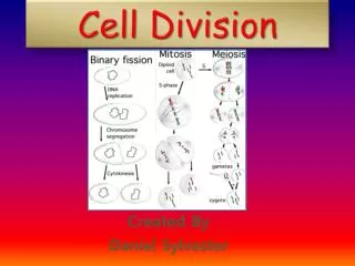
Types of Cell Division • Mitosis • Produces two new daughter cells with the same number and kind of chromosomes as the parent cell • Meiosis • Reduction division produces progeny cells with one-half the genetic content and number of chromosomes as parent cell • Produces gametes/spores
Mitosis • Purpose: • Mitosis occurs in order for organisms to grow and develop. • In order to replenish dead or dying cells such as skin cells, and cells in the digestive tract. • Karyokinesis • process of nuclear division (division of genetic material) • Cytokinesis • Process of dividing cytoplasm/cell
The Cell Cycle • The life of a cell is divided into three stages known as the cell cycle: 1. Interphase: cell carries out normal functions and prepares to divide 2. Mitosis: nucleus divides splits into two 3. Cytokinesis: cell and contents divide into two daughter cells.
Interphase • This phase consist of the G1,S, and G2 phases of the cell cycle. • The chromatin is diffuse. • It may not look like much is going on here, but there is a lot of activity because the cell must prepare for Mitosis: • protein synthesis, • DNA synthesis, • replication of other cellular structures.
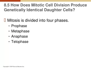
Mitosis • There are 4 main phases: • Prophase, • Metaphase, • Anaphase, • Telophase. • Cytokinesis (division of the cytoplasm) follows and one cell becomes two.
Mitosis: Prophase Major processes during this phase: • Chromosomes condense and form visible bodies • Chromosomes become thicker, shorter, and easily visible when stained under the light microscope. • Two “sister chromatids” join near their middle at a structure called the centromere. • The nucleolus and the nuclear membrane disappear. The mitotic apparatus the spindle, begins to organize within the cell
Mitosis: Metaphase • Chromosomes become aligned at midpoint or equator between poles of the cell • are at their thickest and shortest structure. • They are easily identified as two longitudinally double sister chromatids.
Mitosis: Anaphase • The centromere replicates and splits • The sister chromatids separate and are pulled to opposite sides of the cell
Mitosis: Telophase • Chromosomes now uncoil • Nuclear envelope reappears and surrounds the chromosomes Cytokinesis • The cytoplasm and all its contents are divided between the 2 daughter cells (cytoplasmic division) • membrane creates between the 2 new daughter cells • In plants, such as the onion root tip cells, this is seen as the formation of a cell plate
Mitosis in Root Tip • In a growing plant root, the cells at the tip of the root are constantly dividing to allow the root to grow. • Because each cell divides independently of the others, a root tip contains cells at different stages of the cell cycle. • This makes a root tip an excellent tissue to study the stages of cell division
Materials • Slides & cover slips • Microscope • Fresh onion root tips • Fixative ( methanol-acetic acid 3:1 v/v) • Forceps • 1 M HCl • Razor blade • Stain • Paper towel, or absorbent paper
Method • Cut 2-3 mm of onion root • Use forceps to transfer an onion root tip into the cup of HCl. • Leave for 4 minutes • Transfer the root tip to the cup containing fixative and leave it for 4 minutes. • Then place the root tip on a slide. • Cover the root tip with a few drops of stain for 2 minutes • Cover the root tip with one to two drops of 45% acetic acid • Put a cover slip over the root, put a paper towel or other absorbent paper and with your thumb firmly press on the cover slip.
Observe your preparation under the low power (X10) of a microscope • Search the slide to find cells in various stages of cell division, once you have located cells in division, change to high power (X40) & try to observe several stages of division. • Record the number of cells in each stage. Count at least three full fields of view. You should have counted over 200 cells. • Record your data in the table • Calculate the percentage of cells in each phase and record in the table
Mitotic index • A measure for the proliferation status of a cell population. • It is defined as the ratio between the number ofcellsinmitosisand the total number of cells.
Animation http://www.youtube.com/watch?v=s1ylUTbXyWU
Interphase Prophase Metaphase Anaphase Telophase