Download
1 / 51
510 likes | 634 Views
V alvular heart diseases. Dr. Amanj Kamal F.I.C.M.S. Cardiovascular Surgery. P lease Turn the mobile off. Q uestions at the end. A ortic insufficiency. Result from disease of the valve leaflets or of the aortic root. Etiology. Rheumatic fever Anuloaortic ectasia , as in Marfan .

E N D
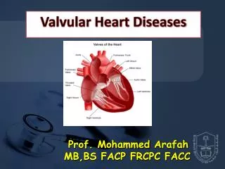
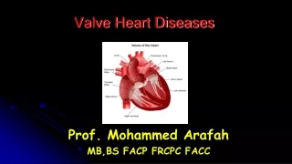
Valvular heart diseases Dr. Amanj Kamal F.I.C.M.S. Cardiovascular Surgery
Aortic insufficiency Result from disease of the valve leaflets or of the aortic root.
Etiology • Rheumatic fever • Anuloaorticectasia, as in Marfan. • Bicuspid. (mostly AS, but AR may occur) • Myxoid degeneration of leaflets. • Infective endocarditis • Trauma • Ascending aortic disection.
Pathophysiology • Diastolic pressure • Coronary blood flow • Work effort of the heart. • (AS: pressure overload, AR: volume overload)
Clinical features • Asymptomatic • Dyspnoea on exertion. • Orthopnoea • Parox. Noctur. Dyspnoea . • Angina • Symptoms of congestive heart failure.
Management • AR requires surgical correction, if medical… • Timing of surgery, difficult in asymptomatic or mild… • Surgery should be done before HF occur • Catheterization needed. • Some times CT-angio, for the ascending and the arch.
Indications of surgery • Symptomatic sever. • Symptomatic acute. • With other diseases (coronary, other valve, aorta..) • Significant LVH • Significant LV dilatation, End Systolic 55 mm • EF less than 30%
Surgical options • Repair of the valve. • Replacement (valve only, or with conduit) • TAVI (Transcatheter Aortic Valve Implantation)
Etiology • Calcific (degenerative). • Congenital • Rheumatic • Other rare causes include • obstructive infective vegetations, • hyperlipoproteinemia, • Paget disease of the bone, • systemic lupus erythematosus, • radiation to the thorax.
Presentation • Patients may present with chest pain, shortness of breath, or syncopal episodes. Physical Exam • Pulse: peak occurs later in systole (pulsustardus) and pulse amplitude is decreased (pulsusparvus). • Murmur: The second characteristic sign is a systolic murmur over the right second intercostal space. The murmur radiates to the carotids . • The second heart sound (S2) is diminished or absent in severe AS
Diagnosis and tests • Electrocardiography: The classic finding is LV hypertrophy. • Chest Radiography: May be normal. However, poststenotic dilatation of the ascending aorta may be evident. Calcification of the aortic valve is rarely seen on radiography but may be seen on fluoroscopy. • Echocardiography: is helpful in assessing the severity of AS, estimating pulmonary systolic pressure; • Cardiac Catheterization: in patients at risk for coronary artery disease prior to valve replacement. • Dobutamine Echocardiography: to assess the severity of the aortic valve lesion
Choices of management • Don’t use vasodilators in AS. • Balloon valvuloplasty. • Surgery (repair or replacement)
Indications of ballooning • Bridge to surgery. In hemodynamically unstable patients. • Palliation for seriously ill patients. • Requiring urgent intervention.
Indications of surgery • Symptomatic (sever or acute), or hypotensive exercise response. • Other surgery needed. • Symptomatic or not but with: • LV hypertrophy • EF less than 50% • valve area less than 1 cm • pressure gradient more than 50 mm Hg
Complications • Congestive heart failure • Sudden cardiac death • Atrial arrhythmias • Infective endocarditis • Systemic calcium embolism • Prosthetic valve related complications
Mitral regurgitation (MR) is the backflow of blood from the left ventricle (LV) to the left atrium (LA) during systole. Patients with mitral regurgitation tolerate pregnancy well, but it might precipitate congestive heart failure in case of severe MR with LV systolic dysfunction.
Etiology • Myxomatous degeneration • Mitral annular calcification • Rheumatic heart disease • Congenital malformation • Ruptured chordaetendineae • Ruptured papillary muscle • Infective endocarditis • Marfan syndrome • Ehlers-Danlos syndrome • Pseudoxanthomaelasticum • Tumors (atrial myxoma) • Functional • Ischemic • Dilated cardiomyopathy • Infiltrative/restrictive cardiomyopathy • Hypertrophic cardiomyopathy • Trauma
Pathophysiology • Severe chronic MR is associated with LVH, LV dilation, increased LVEDP, and heart failure. Symptom onset is related to degree of LA compliance and underlying cause of the MR.
Signs and Symptoms • May remain asymptomatic for many years. • MR secondary to ischemic disease or dilated cardiomyopathy will have symptoms typical of the underlying disease. • The typical initial presentation will be exercise intolerance in the form of exertionaldyspnea. This is followed by symptoms of pulmonary congestion and congestive heart failure (CHF). • The onset of symptoms may coincide with a period of increased hemodynamic burden (e.g., pregnancy, infection, etc.) or with the onset of atrial fibrillation.
Physical Exam • Brisk carotid upstroke with early peak and rapid decline. • displaced diffuse apex late in the course of the disease due to LV dilatation. • Left parasternal pulsations. • Soft S1 with a holosystolic, soft-pitched, and blowing murmur that is loudest at the apex and radiates to the axilla. • A widely split S2
Tests • Electrocardiography • LV hypertrophy in patients with severe MR. • Chest radiograph • LA enlargement later. During the decompensated stage, • Transthoracic echocardiography • Echo Doppler study can estimate both LV and LA volumes/dimensions, LV ejection fraction (LVEF), and the severity of MR, and can help define the anatomic cause of MR. • Transesophageal echocardiography • Useful for evaluation of patients in whom transthoracic echocardiography is inconclusive, • Cardiac catheterization can be performed to assess the severity of MR as well as to delineate coronary anatomy.
management • Medical (anti failure, digoxin, thrombolytics, … • Surgical: • Repair. • Ring anuloplasty • Apparatus sparing replacement. • Replacement.
Indications of surgery • Symptomatic. • Sever structural damages. • Other surgical procedures needed.
Recommendations for coronary angiography in MR: • In patients with angina or history of MI • In patients with one or more risk factors for CAD • When ischemia is suspected as a causal factor in MR
Mitral stenosis (MS) is caused by structural abnormalities in the mitral valve that prevent complete opening during diastole.
Etiology • Rheumatic heart disease is the most common cause of MS. 99% of valves removed for MS have rheumatic involvement. • Other rare causes of MS • Degenerative • Carcinoid syndrome • Whipple's disease • Rheumatoid arthritis • Obstruction by large vegetation • Congenital mitral stenosis accounts for <1% of cases.
management • Medical (anti failure, digoxin, thrombolytics, … • Surgical: • Balloon anuloplasty • Repair. Comisurotomy • Apparatus sparing replacement. • Replacement.
Consederations For all valves
Child bearing age • Time of intervention. • Termination of pregnancy or not. • Type of anasthesia • Use of anticoagulants. • Choice of the valve. • How frequent to follow up.
Choice of the valve For all valvular diseases
Depend on: • Age of the patient. • Child bearing age. • Patient away from hospital • Contraindications to anticoagulants • Position of the valve. • Use of anticoagulants for other purposes.
Mechanical valves: • Ball and Socket (Starr Edward) • Tilting disc (Shiely, medtronic) • Bileaflets (St Jude, Carbomedic, Duromedic) • Tissue valves: • Porcine (Carpentire Edward, Hancock, Medtronic intact) • Porcine stentless (Torento, carpentire Edward stentless, ..) • Pericardial (Carpentire Edward Bovine) • Homograft (Cadaver) • Xenograft • Autograft.
Single tilting leaflet St. Jude Ball and socket Shiely