Download
1 / 76
880 likes | 1.34k Views
Valvular Heart Diseases . Prof. Mohammed Arafah MB,BS FACP FRCPC FACC. ALL cardiac valves can be involved in pathological processes . Etiology. Congenital : - Bicuspid or unicuspid . - Subvalvular or supravalvular . . Etiology - continue.

E N D
Valvular Heart Diseases Prof. Mohammed Arafah MB,BS FACP FRCPC FACC
ALL cardiac valves can be involved in pathological processes
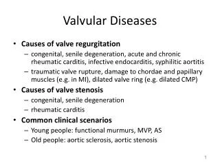
Etiology • Congenital : - Bicuspid or unicuspid . - Subvalvular or supravalvular .
Etiology - continue • Acquired : - Rheumatic . - Degeneration . - myxomatous - calcification - Ischaemic . - Infective Endocarditis . - Valve ring dilatation .
TYPES of Presentations • Acute Presentation : - Acute mitral regurgitation due to eg acute myocardial infarction acute chordeatendineae rupture
TYPES of Presentations • Chronic Presentation : - Chronic mitral regurgitation due to eg RHRUMATIC fever . Mitral valve Prolapse . - Chronic aortic regurgitation due to eg Bicuspid Aortic valve .
HEAMODYNAMICS Consequences • Pressure Overload : - Aortic stenosis Left Ventricular hypertrophy - Mitral stenosis Left Atriarl hypertrophy & dilatation
HEAMODYNAMICS Consequences • Volume Overload : - chronic mitral regurgitation dilated left ventricle & left atria - chronic tricuspid regurgitation dilated right ventricle & right atria
SYMPTOMS • Dyspnea , paroxysmal nocturnal dyspnea orthopnea . • Palpitation . • Chest pain . • Dizziness , prefainting ,syncope . • Oedema , Ascites • Cough . • Fatigue • Hemoptysis • Symptoms of thromboembolic complication .
Signs of Valvular Diseases • Abnormal look ( mitral facies ) . • Abnormal pulse ( Atrial fibrillation ) . • Abnormal JVP • Apex beat abnormality . • Sternal or parasternal heave . • Thrill . • Abnormal heart sound . • MURMURS . Systolic or Diastolic .
INVESTIGATION • ECG . • CXR . • Echo cardiology . M mode , 2D ,3D . 4 D . TEE . Doppler . • 24 hours monitor for heart rhythm . • MRI . • Cardiac catheterization .
ETIOLOGY Rheumatic Fever which is related to streptococcus infections, causing damage to the mitral valve and leading to mitral stenosis later in life.
OTHER LESS COMMON CAUSES OF MITRAL STENOSIS Congenital Mitral Stenosis Systemic Lupus Erythematosus Rheumatoid Arthritis AtrialMyxoma Malignant Carcinoid Bacterial Endocarditis
MITRAL STENOSISresults in several changes to the integrity of the valves: CUSPS THICKEN COMMISSURES FUSED TOGETHER CHORDAE TENDINAE BECOMES THICKENED & SHORTENED CALCIUM DEPOSITS FORM
SIGNS & SYMPTOMS • Symptoms of mitral stenosis usually begin with the hallmark signs of DYSPNEA ON EXERTION! • The first bouts of dyspnea in patients with mitral stenosis are usually precipitated by exercise, emotional stress, infection, or atrial fibrillation, all of which increase the rate of blood flow across the mitral orifice & result in further elevation of Left
OTHER PRINCIPAL SIGNS AND SYMPTOMS INCLUDES: • Fatigue • Orthopnea • Paroxysmal nocturnal dyspnea • Pulmonary edema – develops when there’s a sudden ↑ in flow rate across a markedly narrowed mitral orifice. • Palpitations – owing to presence of arrhythmias • Hemoptysis – due to rupture of thin dilated bronchial veins. • Peripheral edema .
The Diagnostic testing used to evaluate the presence & severity of Mitral Stenosis includes: ECG Chest Radiograph 2D Echocardiogram Doppler Study TransEsophageal Echocardiography
LEFT PARASTERNAL, LONG AXIS VIEW STENOTIC MITRAL VALVE
COMPLICATIONS OF MITRAL STENOSIS ATRIAL FIBRILLATION LUNG CONGESTION BLOOD CLOTS with SYSTEMIC EMBOLIZATION PULMOARY HYPERTENSION CONGESTIVE HEART FAILURE
MEDICAL MANAGEMENT DIURETICS DIGITALIS ANTI-ARRYHTHMICS ANTICOAGULANTS ANTIBIOTICS
Intervention • PERCUTANEOUS TRANSVENOUS MITRAL COMMISSUROTOMY (PTMC) • SURGICAL COMMISSUROTOMY • MITRAL VALVE Replacement .
MVA = .982 cm² MVA = 1.84 cm² PRE-PROCEDURE POST-PROCEDURE
ETIOLOGY • RHEUMATIC HEART disease . • MITRAL Valve Prolapse . • Others - IHD - Cardiomyopathy ( dilated , hypertrophic ) - Hypertensive heart disease - infective endocarditis - Myocarditis - connective tissue disorders - (SLE) -collagen abnormalities - Marfan's syndrome
SIGNS • Laterally displaced (forceful) diffuse apex beat and a systolic thrill . • Soft first heart sound . • Pansystolic murmur . • Prominent third heart sound .
Management of mitral regurgitation • Evidence of progressive cardiac enlargement generally warrants early surgical intervention by either mitral valve repair or replacement . • Treatment with ACE inhibitors, diuretics and possibly anticoagulants .
Pathology • Large mitral valve leaflets, an enlarged mitral annulus, abnormally long chordae or disordered papillary muscle contraction . • Demonstrate myxomatous degeneration of the mitral valve leaflets . • Associated with Marfan's syndrome, thyrotoxicosis, rheumatic or ischaemic heart disease .