Download
1 / 54
540 likes | 564 Views
Discover how sensory receptors transduce stimuli, sensations become action potentials, and perceptions form in the brain through sensory reception. Explore the functions performed by sensory receptors and types of receptors based on energy transduction.

E N D
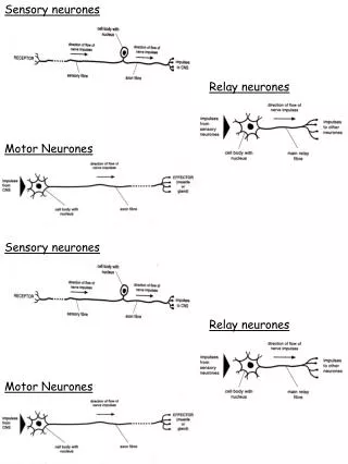
Sensory receptors transduce stimulus energy and transmit signals to the central nervous system • Sensations are action potentials • That reach the brain via sensory neurons • Once the brain is aware of sensations • It interprets them, giving the perception of stimuli
Sensations and perceptions • Begin with sensory reception, the detection of stimuli by sensory receptors • Exteroreceptors • Detect stimuli coming from the outside of the body • Interoreceptors • Detect internal stimuli
Functions Performed by Sensory Receptors • All stimuli represent forms of energy • Sensation involves converting this energy • Into a change in the membrane potential of sensory receptors • Sensory receptors perform four functions in this process • Sensory transduction, amplification, transmission, and integration
Weakmuscle stretch Strongmuscle stretch Muscle Dendrites Receptor potential –50 –50 –70 –70 Stretchreceptor Membranepotential (mV) Action potentials 0 0 Axon –70 –70 4 5 1 2 6 7 0 0 1 2 3 4 5 6 7 3 Time (sec) Time (sec) stretch, producing a receptor potential in the stretch receptor. The receptor potential triggers action potentials in the axon of the stretch receptor. A stronger stretch producesa larger receptor potential and higherrequency of action potentials. (a) Crayfish stretch receptors have dendrites embedded in abdominal muscles. When the abdomen bends, muscles and dendrites Figure 49.2a • Two types of sensory receptors exhibit these functions • A stretch receptor in a crayfish
Fluid moving inone direction Fluid moving in other direction No fluidmovement “Hairs” ofhair cell Neuro-trans-mitter at synapse Moreneuro-trans-mitter Lessneuro-trans-mitter –50 –50 Axon –50 Receptor potential –70 –70 –70 Membranepotential (mV) Membranepotential (mV) Membranepotential (mV) Action potentials 0 0 0 –70 –70 –70 5 6 7 1 2 3 4 0 0 0 1 2 3 4 5 6 7 1 2 3 4 5 6 7 Time (sec) Time (sec) Time (sec) (b) Vertebrate hair cells have specialized cilia or microvilli (“hairs”) that bend when sur-rounding fluid moves. Each hair cell releases an excitatory neurotransmitter at a synapse with a sensory neuron, which conducts action potentials to the CNS. Bending in one direction depolarizes the hair cell, causing it to release more neurotransmitter and increasing frequency Figure 49.2b • A hair cell found in vertebrates of action potentials in the sensory neuron. Bending in the other direction has the opposite effects. Thus, hair cells respond to the direction of motion as well as to its strength and speed.s
Types of Sensory Receptors • Based on the energy they transduce, sensory receptors fall into five categories • Mechanoreceptors • Chemoreceptors • Electromagnetic receptors • Thermoreceptors • Pain receptors
Cold Light touch Pain Hair Heat Epidermis Dermis Hair movement Nerve Strong pressure Connective tissue • The mammalian sense of touch • Relies on mechanoreceptors that are the dendrites of sensory neurons Figure 49.3
Some snakes have very sensitive infrared receptors • That detect body heat of prey against a colder background Figure 49.5a (a) This rattlesnake and other pit vipers have a pair of infrared receptors,one between each eye and nostril. The organs are sensitive enoughto detect the infrared radiation emitted by a warm mouse a meter away. The snake moves its head from side to side until the radiation is detected equally by the two receptors, indicating that the mouse is straight ahead.
Many mammals appear to use the Earth’s magnetic field lines • To orient themselves as they migrate Figure 49.5b (b) Some migrating animals, such as these beluga whales, apparentlysense Earth’s magnetic field and use the information, along with other cues, for orientation.
The mechanoreceptors involved with hearing and equilibrium detect settling particles or moving fluid • Hearing and the perception of body equilibrium • Are related in most animals
Tympanicmembrane 1 mm • Many arthropods sense sounds with body hairs that vibrate • Or with localized “ears” consisting of a tympanic membrane and receptor cells Figure 49.7
1 2 Overview of ear structure The middle ear and inner ear Incus Semicircularcanals Skullbones Stapes Middleear Outer ear Inner ear Malleus Auditory nerve,to brain Pinna Tympanicmembrane Cochlea Eustachian tube Auditory canal Ovalwindow Eustachian tube Tympanicmembrane Tectorialmembrane Hair cells Roundwindow Cochlear duct Bone Vestibular canal Auditory nerve Axons of sensory neurons Basilarmembrane To auditorynerve Tympanic canal Organ of Corti 4 3 The organ of Corti The cochlea • Exploring the structure of the human ear Figure 49.8
Hearing • Vibrating objects create percussion waves in the air • That cause the tympanic membrane to vibrate • The three bones of the middle ear • Transmit the vibrations to the oval window on the cochlea
Cochlea Stapes Axons ofsensoryneurons Oval window Vestibularcanal Perilymph Apex Base Roundwindow Tympaniccanal Basilar membrane • These vibrations create pressure waves in the fluid in the cochlea • That travel through the vestibular canal and ultimately strike the round window Figure 49.9
The pressure waves in the vestibular canal • Cause the basilar membrane to vibrate up and down causing its hair cells to bend • The bending of the hair cells depolarizes their membranes • Sending action potentials that travel via the auditory nerve to the brain
Lateralline Opening of lateralline canal Lateral line canal Scale Epidermis Neuromast Lateral nerve Segmental muscles of body wall Cupula Sensoryhairs Supporting cell Hair cell Nerve fiber • The lateral line system contains mechanoreceptors • With hair cells that respond to water movement Figure 49.12
The senses of taste and smell are closely related in most animals • The perceptions of gustation (taste) and olfaction (smell) • Are both dependent on chemoreceptors that detect specific chemicals in the environment
Taste in Humans • The receptor cells for taste in humans • Are modified epithelial cells organized into taste buds • Five taste perceptions involve several signal transduction mechanisms • Sweet, sour, salty, bitter, and umami (elicited by glutamate)
Taste pore Sugar molecule Taste bud Sensoryreceptorcells Sensoryneuron Tongue 1 A sugar molecule binds to a receptor protein on the sensory receptor cell. Sugar Adenylyl cyclase G protein Sugarreceptor 2Binding initiates a signal transduction pathway involving cyclic AMP and protein kinase A. ATP cAMP Proteinkinase A 3Activated protein kinase A closes K+ channels in the membrane. SENSORYRECEPTORCELL K+ 4 The decrease in the membrane’s permeability to K+ depolarizes the membrane. Synapticvesicle —Ca2+ 5 Depolarization opens voltage-gated calcium ion (Ca2+) channels, and Ca2+ diffuses into the receptor cell. Neurotransmitter 6 The increased Ca2+ concentration causes synaptic vesicles to release neurotransmitter. Sensory neuron • Transduction in taste receptors • Occurs by several mechanisms Figure 49.14
Brain Action potentials Odorant Olfactory bulb Nasal cavity Bone Epithelial cell Odorantreceptors Chemoreceptor Plasmamembrane Cilia Figure 49.15 Odorant Mucus • When odorant molecules bind to specific olfactory receptors • A signal transduction pathway is triggered, sending action potentials to the brain
Similar mechanisms underlie vision throughout the animal kingdom • Many types of light detectors • Have evolved in the animal kingdom and may be homologous
Two major types of image-forming eyes have evolved in invertebrates • The compound eye and the single-lens eye
(a) The faceted eyes on the head of a fly, photographed with a stereomicroscope. 2 mm (b) The cornea and crystalline cone of each ommatidium function as a lens that focuses light on the rhabdom, a stack of pigmented plates inside a circle of photoreceptors. The rhabdom traps light and guides it to photoreceptors. The image formed by a compound eye is a mosaic of dots produced by different intensities of light entering the many ommatidia from different angles. Cornea Lens Crystallinecone Rhabdom Photoreceptor Axons Ommatidium • Compound eyes are found in insects and crustaceans • And consist of up to several thousand light detectors called ommatidia Figure 49.17a–b
Single-lens eyes • Are found in some jellies, polychaetes, spiders, and many molluscs • Work on a camera-like principle
Structure of the Eye • The main parts of the vertebrate eye are • The sclera, which includes the cornea • The choroid, a pigmented layer • The conjunctiva, that covers the outer surface of the sclera • The iris, which regulates the pupil • The retina, which contains photoreceptors • The lens, which focuses light on the retina
Sclera Choroid Retina Ciliary body Fovea (centerof visual field) Suspensoryligament Cornea Iris Opticnerve Pupil Aqueoushumor Lens Vitreous humor Central artery and vein of the retina Optic disk(blind spot) • The structure of the vertebrate eye Figure 49.18
Front view of lensand ciliary muscle Ciliary muscles contract, pullingborder of choroid toward lens Lens (rounder) Choroid Retina Suspensory ligaments relax Ciliarymuscle Lens becomes thicker and rounder, focusing on near objects Suspensoryligaments (a) Near vision (accommodation) Ciliary muscles relax, and border of choroid moves away from lens Lens (flatter) Suspensory ligaments pull against lens Lens becomes flatter, focusing on distant objects (b) Distance vision • Humans and other mammals • Focus light by changing the shape of the lens Figure 49.19a–b
The human retina contains two types of photoreceptors • Rods are sensitive to light but do not distinguish colors • Cones distinguish colors but are not as sensitive
Muscles move skeletal parts by contracting • The action of a muscle • Is always to contract
Human Grasshopper Extensormusclerelaxes Bicepscontracts Tibiaflexes Flexormusclecontracts Tricepsrelaxes Forearmflexes Extensormusclecontracts Tibiaextends Bicepsrelaxes Forearmextends Flexormusclerelaxes Triceps contracts • Skeletal muscles are attached to the skeleton in antagonistic pairs • With each member of the pair working against each other Figure 49.27
Muscle Bundle ofmuscle fibers Nuclei Single muscle fiber (cell) Plasma membrane Myofibril Z line Lightband Dark band Sarcomere TEM 0.5 m A band I band I band M line Thickfilaments(myosin) Thinfilaments(actin) H zone Z line Z line Sarcomere Vertebrate Skeletal Muscle • Vertebrate skeletal muscle • Is characterized by a hierarchy of smaller and smaller units Figure 49.28
A skeletal muscle consists of a bundle of long fibers • Running parallel to the length of the muscle • A muscle fiber • Is itself a bundle of smaller myofibrils arranged longitudinally
The myofibrils are composed to two kinds of myofilaments • Thin filaments, consisting of two strands of actin and one strand of regulatory protein • Thick filaments, staggered arrays of myosin molecules • Skeletal muscle is also called striated muscle • Because the regular arrangement of the myofilaments creates a pattern of light and dark bands
Each repeating unit is a sarcomere • Bordered by Z lines • The areas that contain the myofilments • Are the I band, A band, and H zone
The Sliding-Filament Model of Muscle Contraction • According to the sliding-filament model of muscle contraction • The filaments slide past each other longitudinally, producing more overlap between the thin and thick filaments
0.5 m (a) Relaxed muscle fiber. In a relaxed muscle fiber, the I bandsand H zone are relatively wide. Z H A Sarcomere (b) Contracting muscle fiber. During contraction, the thick andthin filaments slide past each other, reducing the width of theI bands and H zone and shortening the sarcomere. (c) Fully contracted muscle fiber. In a fully contracted musclefiber, the sarcomere is shorter still. The thin filaments overlap,eliminating the H zone. The I bands disappear as the ends ofthe thick filaments contact the Z lines. • As a result of this sliding • The I band and the H zone shrink Figure 49.29a–c
The sliding of filaments is based on • The interaction between the actin and myosin molecules of the thick and thin filaments • The “head” of a myosin molecule binds to an actin filament • Forming a cross-bridge and pulling the thin filament toward the center of the sarcomere
Thick filament Thin filaments 1 Starting here, the myosin head is bound to ATP and is in its low-energy confinguration. 5 Binding of a new mole- cule of ATP releases the myosin head from actin, and a new cycle begins. Thin filament Myosin head (low-energy configuration) The myosin head hydrolyzes ATP to ADP and inorganic phosphate ( I ) and is in its high-energy configuration. ATP 2 ATP Cross-bridge binding site Thick filament P Actin Thin filament moves toward center of sarcomere. Myosin head (high-energy configuration) ADP Myosin head (low-energy configuration) P i 1 The myosin head binds toactin, forming a cross-bridge. 3 ADP + Cross-bridge ADP P i P i Releasing ADP and ( i), myosinrelaxes to its low-energy configuration, sliding the thin filament. 4 P • Myosin-actin interactions underlying muscle fiber contraction Figure 49.30
Tropomyosin Ca2+-binding sites Actin Troponin complex (a) Myosin-binding sites blocked • When a muscle is at rest • The myosin-binding sites on the thin filament are blocked by the regulatory protein tropomyosin Figure 49.31a
Ca2+ Myosin-binding site (b) Myosin-binding sites exposed • For a muscle fiber to contract • The myosin-binding sites must be uncovered • This occurs when calcium ions (Ca2+) • Bind to another set of regulatory proteins, the troponin complex Figure 49.31b
Motorneuron axon Mitochondrion Synapticterminal T tubule Sarcoplasmicreticulum Ca2+ releasedfrom sarcoplasmicreticulum Myofibril Sarcomere Plasma membraneof muscle fiber • The stimulus leading to the contraction of a skeletal muscle fiber • Is an action potential in a motor neuron that makes a synapse with the muscle fiber Figure 49.32
The synaptic terminal of the motor neuron • Releases the neurotransmitter acetylcholine, depolarizing the muscle and causing it to produce an action potential
Action potentials travel to the interior of the muscle fiber • Along infoldings of the plasma membrane called transverse (T) tubules • The action potential along the T tubules • Causes the sarcoplasmic reticulum to release Ca2+ • The Ca2+ binds to the troponin-tropomyosin complex on the thin filaments • Exposing the myosin-binding sites and allowing the cross-bridge cycle to proceed
Acetylcholine (ACh) released by synaptic terminal diffuses across synapticcleft and binds to receptor proteins on muscle fiber’s plasma membrane, triggering an action potential in muscle fiber. Synapticterminalof motorneuron 1 PLASMA MEMBRANE Synaptic cleft T TUBULE Action potential is propa- gated along plasma membrane and down T tubules. 2 ACh SR 4 Action potential triggers Ca2+ release from sarco- plasmic reticulum (SR). 3 Ca2 Calcium ions bind to troponin; troponin changes shape, removing blocking action of tropomyosin; myosin-binding sites exposed. Tropomyosin blockage of myosin- binding sites is restored; contraction ends, and muscle fiber relaxes. 7 Ca2 CYTOSOL Cytosolic Ca2+ is removed by active transport into SR after action potential ends. 6 ADP P2 Myosin cross-bridges alternately attach to actin and detach, pulling actin filaments toward center of sarcomere; ATP powers sliding of filaments. 5 • Review of contraction in a skeletal muscle fiber Figure 49.33
Neural Control of Muscle Tension • Contraction of a whole muscle is graded • Which means that we can voluntarily alter the extent and strength of its contraction • There are two basic mechanisms by which the nervous system produces graded contractions of whole muscles • By varying the number of fibers that contract • By varying the rate at which muscle fibers are stimulated
Motorunit 1 Motorunit 2 Spinal cord Synaptic terminals Nerve Motor neuroncell body Motor neuronaxon Muscle Muscle fibers Tendon • In a vertebrate skeletal muscle • Each branched muscle fiber is innervated by only one motor neuron • Each motor neuron • May synapse with multiple muscle fibers Figure 49.34
A motor unit • Consists of a single motor neuron and all the muscle fibers it controls • Recruitment of multiple motor neurons • Results in stronger contractions
Tetanus Tension Summation of two twitches Singletwitch Time Actionpotential Pair ofactionpotentials Series of action potentials at high frequency • A twitch • Results from a single action potential in a motor neuron • More rapidly delivered action potentials • Produce a graded contraction by summation Figure 49.35
Tetanus is a state of smooth and sustained contraction • Produced when motor neurons deliver a volley of action potentials