Download
1 / 145
1.45k likes | 1.82k Views
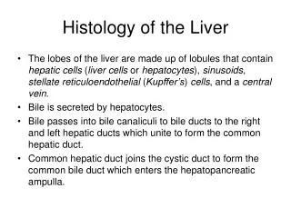
HISTOLOGY OF THE LIVER. HEPATIC LOBULES vs. ACINI. Portal tract. 3. 2. 1. Central vein. Portal tract. Acini are defined by micro- circulatory layout of liver, with a central axis (portal tract & its afferent vessels), surrounded by 3 zones.

E N D
HISTOLOGY OF THELIVER • HEPATIC LOBULES vs. ACINI Portal tract 3 2 1 Central vein Portal tract Acini are defined by micro- circulatory layout of liver, with a central axis (portal tract & its afferent vessels), surrounded by 3 zones Traditional conception of liver histology, arbitrarily divided into centrilobular, periportal (peripheral) & midlobular zones
Portal triads: contain a bile duct, a small hepatic artery and a portal vein branch, surrounded by type I & III collagen Central vein (tributary of hepatic vein) with blood to hepatic parenchyma flowing from the portal triads to the central veins HISTOLOGY OF THELIVER
DISEASES OF THELIVER • Hepatic injury • Jaundice & cholestasis • Hepatic failure • Cirrhosis • Inflammatory disorders: hepatitis, abscesses • Drug & toxin-related diseases: alcohol liver disease • Inborn error of metabolism & pediatric liver disease: Hemochromatosis, Wilson’s disease, neonatal hepatitis & Reye’s syndrome • Intrahepatic biliary tract disease: PBC, PSC • Circulatory disorders • Tumors
HISTOLOGIC PATTERNS OFHEPATIC INJURY • Inflammation: acute or chronic hepatitis; portal or lobular • Degeneration: ballooning, foamy, steatosis • Necrosis: coagulative or lytic (hydropic); Councilman bodies; centrilobular, focal, piece-meal, bridging, submassive, massive • Fibrosis: portal, central, bridging • Cirrhosis: regenerative nodules surrounded by fibrosis
Fat (neutral fat, triglycerides) in liver cells indicates defect in lipid metabolism or lipoprotein synthesis or unusual amounts of adipose or dietary lipids brought to liver HISTOLOGIC PATTERNS OF HEPATIC INJURYSTEATOSIS
Swelling or hydropic change is a result of defects in membrane and/or mitochondrial function HISTOLOGIC PATTERNS OF HEPATIC INJURYHEPATOCYTE SWELLING
Coagulative necrosis: poorly staining mummified hepatocytes Councilman bodies: dead hepatocytes Lytic necrosis: hepatocytes swell & rupture Histologic patterns of hepatic injuryNECROSIS
PHYSIOLOGY OF THE LIVER BILIRUBIN METABOLISM MACROPHAGE • Aging RBCsHEME Heme oxygenase BILIVERDIN Biliverdin reductase beta-glucuronidase • BILIRUBIN- • Albumin complex U R O B I L I N O G E N BILIRUBIN GLUCORONIDES UDP
PHYSIOLOGY OF THE LIVER • In addition to bilirubin, the liver secretes 12-36 g bile acids/day: carboxylated steroid molecules derived from cholesterol & hydroxyl groups • Cholic acid & chenodeoxycholic acid • Secreted as taurine & glycine conjugates • 10-20% are deconjugated in ileum • 0.2-0.6 g/d fecal loss matched by de novo liver synthesis • Functions of hepatic bile: • 1) Primary pathway for elimination of water-insoluble bilirubin, excess cholesterol & xenobiotics • 2) Emulsification of dietary fat in gut lumen
PATHOLOGY OF THE LIVERJAUNDICE • Jaundice: yellowish discoloration of skin & sclera (icterus) due to systemic retention of bilirubin (> 2 mg/dl) • Equilibrium between bilirubin production & clearance is disturbed: • 1) Excessive production • 2) Reduced hepatocellular uptake • 3) Impaired conjugation • 4) Decreased hepatocellular excretion • 5) Impaired bile flow • Kernicterus: accumulation of bilirubin in brain
UNCONJUGATED BILIRUBIN Water-insoluble Tightly complexed to serum albumin Cannot be excreted in urine Free form is toxic Lab test: Total bilirubin minus direct bilirubin CONJUGATED BILIRUBIN Water-soluble Loosely bound to serum albumin Excess amounts are excreted in urine Nontoxic Lab test: measured by direct bilirubin TYPES OFJAUNDICE
LAB EVALUATION OF LIVER DISEASELIVER FUNCTION TESTS • Tests of hepatocyte integrity • ASL (SGOT)* • ALT (SGPT)* • LDH • Tests of biliary excretory function • Serum Bilirubin* • Alkaline phosphatase* • Gamma-glutamyl transpeptidas • Tests of hepatocyte function • Albumin* • Prothrombinetime* • Ammonia • Aminopyrine breath test; galactose elimination
PATHOLOGY OF THE LIVERCHOLESTASIS • Systemic retention of bilirubin and other solutes eliminated in bile (e.g. bile salts & cholesterol) • Results from hepatocellular dysfunction & (intra- or extrahepatic) biliary obstruction • c/o: Jaundice, pruritis, skin xanthomas, malabs. • Lab: Elevated bilirubin, alk. phosphatase, lipids • Bx: bile pigment accumulation, foamy degenerat- ion, bile duct distension & proliferation, bile lakes, portal tract fibrosis, cholangitis & cholangiolitis • Types: • Intrahepatic • Extrahepatic
PATHOLOGY OFCHOLESTASIS • Accumulation of bile pigment within hepatic parenchyma • Hepatocyte swelling and foamy degeneration • Bile duct proliferation secondary to biliary tree obstruction • Hepatocyte necrosis, bile lakes, & portal tract fibrosis
PEDIATRIC LIVER DISEASESNEONATAL CHOLESTASIS • Prolonged conjugated hyperbilirubinemia in the newborn • Major causes: EHBA & neonatal hepatitis • Bile duct obstruction: Extrahepatic biliary atresia (EHBA) • Neonatal infections: CMV, sepsis, UTI, syphilis • Toxic: drugs, parenteral nutrition • Metabolic diseases: tyrosinemia, Niemann-Pick disease, galactosemia, AAT deficiency, cystic fibrosis, .. • Miscellaneous: shock, hypoperfusion, Alagille’s syndrome (paucity of bile ducts), .. • Idiopathic neonatal hepatitis
PEDIATRIC LIVER DISEASESNEONATAL CHOLESTASIS • Clinical presentation is typical: jaundice, dark urine, light stools, hepatomegaly • Neonatal hepatitis may be primary (idiopathic) or secondary • Idiopathic neonatal hepatitis (50-60%), extrahepatic biliary atresia (20%) & AAT deficiency (15%) • Distinction between these disorders is essential because management is different • Liver biopsy is important in the diagnosis
PATHOLOGY OF THE LIVERHEPATIC FAILURE • Mostly due to progressive, or less often sudden massive hepatic destruction with loss of 80-90% of hepatic functional capacity • Causes: • 1) Chronic liver diseases. • 2) Massive hepatic necrosis (fulminant failure): viral hepatitis, drug & chemical toxicity (acetaminophen, halothane, rifampicin, MOI antidepressants, CCl4, Amanita mushroom toxins • 3) Hepatic dysfunction without overt necrosis: viable but nonfunctional hepatocytes, e.g. Reye’s syndrome, tetracycline toxicity, acute fatty liver of pregnancy
Table 18-2. Clinical Consequences of Liver Disease Characteristic signs • Hepatic dysfunction: • Jaundice and cholestasis • Hypoalbuminemia • Hyperammonemia • Hypoglycemia • Fetor hepaticus • Palmar erythema • Spider angiomas • Hypogonadism • Gynecomastia • Weight loss • Muscle wasting • Portal hypertension from cirrhosis: • Ascites • Splenomegaly • Hemorrhoids • Caput medusae-abdominal skin
Life-threatening complications • Hepatic failure • Multiple organ failure • Coagulopathy • Hepatic encephalopathy • Hepatorenal syndrome • Portal hypertension from cirrhosis • Esophageal varices, risk of rupture • Malignancy with chronic disease • Hepatocellular carcinoma
PATHOLOGY OF THE LIVERHEPATIC FAILURE • Most cases are due to overwhelming viral hepatitis and alcoholic liver disease • Symptoms may occur within days with or without prior history of liver disease • A variety of stressful events may contribute to onset of failure: • GI bleeding • Acute infections • Electrolyte disturbances • Major surgery, heart failure, shock • Treatment: not satisfactory • Px: 80% mortality rate
PATHOLOGY OF THE LIVERHEPATIC ENCEPHALOPATHY • A metabolic disorder of CNS & neuromuscular system associated with severe loss of hepatocellular function & portosystemic shunting • The brain is exposed to an altered metabolic environment (ammonia?) which impairs neuronal function & promotes generalized brain edema • Patients exhibit a wide range of disturbances of consciousness: subtle behavioural changes, confusion, stupor, deep coma & death • Other neurologic signs: Rigidity, hyperreflexia, EEG changes, seizures, asterixis • Minor morphologic changes in brain
PATHOLOGY OF THE LIVERHEPATO-RENAL SYNDROME • Development of renal failure in patients with severe liver disease, without presence of intrinsic morphologic or functional causes in the kidney • Excluded are cases of concomitant damage to liver & kidneys and acute tubular necrosis secondary to circulatory collapse • Pathogenesis: due to VC & decreased renal BF? • c/o: decrease in urine output • Retained ability to concentrate urine: Hyperosmolar urine, protein -ve & low Na+ • Lab: Increased blood urea and creatinine • Px: May hasten death or may persist for months
PATHOLOGY OF THE LIVERCIRRHOSIS • Irreversible end stage of chronic liver disease, which leads to parenchymal injury and fibrosis • 3 histologic features: • 1) Bridging fibrous septa • 2) Disruption of entire liver architecture • 3) Parenchymal nodules
ETIOLOGIC CLASSIFICATION OFCIRRHOSIS • Viral hepatitis • Alcoholic liver disease • Biliary diseases • Genetic hemochromatosis • Wilson’s disease • a1-antitrypsin deficiency • Drugs (a-methyldopa, acetaminophen…) • Ca, syphilis • Galactosemia, tyrosinosis.. • “Cardiac cirrhosis” • Cryptogenic cirrhosis
Alcoholic liver disease 60% to 70% Viral hepatitis 10% Biliary diseases 5% to 10% Primary hemochromatosis 5% Wilson disease Rare α1-Antitrypsin deficiency Rare Cryptogenic cirrhosis 10% to 15% Etiologic Factors Of Cirrhosis
PATHOGENESIS OF CIRRHOSIS • Progressive fibrosis with collagen types I & III deposited in all portions of lobule. (Ito cell) • Collagen synthesis & deposition is stimulated by: • Chronic inflammation & cytokine production (TNF, IL-1) • Cytokine production by endogenous liver cells • Disruption of extracellular matrix • Direct stimulation of Ito cells by toxins • This is accompanied by alterations in sinusoidal endothelial cells • > resulting in severe disruption of blood flow & impaired diffusion of solutes & proteins
Micronodular cirrhosis nodules & scars of uniform size Macronodular cirrhosis nodules & scars of variable size PATHOLOGY OFCIRRHOSIS
CLINICAL FEATURES OFCIRRHOSIS • Clinical features: • May be asymptomatic • Nonspecific symptoms: malaise, anorexia, weight loss, weakness • Jaundice, ascites, peripheral edema • serum transaminases, bilirubin, alk. phosph. • serum protein (globulins, albumin, C.F.), Hb • Advanced disease: frank debilitation • Hepatic failure • Prognosis:ultimately will die of: • Progressive hepatic failure • Portal hypertension • Hepatocellular carcinoma
PATHOLOGY OF THE LIVERPORTAL HYPERTENSION • = Increased resistance to portal blood flow • Causes: • I. Pre-hepatic: • Portal vein thrombosis • Massive splenomegaly • II. Hepatic: • Cirrhosis • III. Post-hepatic: • Severe right-sided heart failure • Constrictive pericarditis • Hepatic vein outflow obstruction
LIVER CIRRHOSIS CAUSINGPORTAL HYPERTENSION • Due to: • Increased resistance to portal blood flow in sinusoids • Compression of central veins by perivenular fibrosis & expanding parenchymal nodules • Anastomoses between arterial & portal systems in fibrous bands • Consequences: • 1) Ascites • 2) Portosystemic venous shunts • 3) Congestive splenomegaly • 4) Hepatic encephalopathy
COMPLICATIONS OF PORTAL HYPERTENSIONASCITES • Excess (usually serous) fluid in abdominal cavity • Fluid: albumin, solutes, few mesothelial cells, mononuclear leukocytes • Neutrophils=secondary infection • Hydrothorax • Pathogenesis: • Sinusoidal hypertension: increased hydrostatic pressure & hypoalbuminemia • Percolation of hepatic lymph into peritoneal cavity • Renal retention of sodium & water secondary to hyperaldosteronism
COMPLICATIONS OF PORTALHYPERTENSIONPORTO-SYSTEMIC SHUNTS • Bypasses develop wherever the systemic & portal circulation share capillary beds • Sites: • Rectum • Cardio-esophageal junction • Retroperitoneum • Falciform ligament of liver (periumbilical & abdominal wall)
COMPLICATIONS OF PORTALHYPERTENSIONSPLENOMEGALY • Congestive splenomegaly • May be massive • Hypersplenism: removal of excessive amounts of one or more of the formed blood elements: • Anemia • Leukopenia • Thrombocytopenia
INFLAMMATIONS OF THE LIVERINFECTIONS • Pathways: • Arterial supply • Portal vein • Ascending infection in the biliary tract • Direct liver invasion from nearby infection • Penetrating injury • Organisms: • Bacterial: Staphylococcal bacteremia, salmonelloses, miliary TB • Viral: infectious mononucleosis • Protozoal: malaria, amebiasis, echinococcus
INFLAMMATIONS OF THE LIVERVIRAL HEPATITIS • EBV • CMV • HSV • Arbovirus (Yellow fever) • Rubella • Adenovirus • Enterovirus • Hepatotropic (Hepatitis) viruses: A, B, C, D, E, G
CLINICAL SYNDROMES OFVIRAL HEPATITIS • Carrier state • Asymptomatic infection • Acute hepatitis • Chronic hepatitis • Fulminant hepatitis
CLINICAL SYNDROMES OF VIRAL HEPATITISCARRIER STATE • Reservoir of infection • “healthy” carrier • asymptomatic chronic liver disease • essentially normal liver biopsy • HBV: • 90-95% of early life infections; vertical transmission • 1-10% of infections in adulthood • individuals with impaired immunity • “ground-glass” hepatocytes, “sanded” nuclei • HCV: • 0.2-1% of blood donors • chronic hepatitis usually present histologically • HDV: • 1-10% of drug addicts; in conjunction with HBV
CLINICAL SYNDROMES OF VIRAL HEPATITISASYMPTOMATIC INFECTION • Serologic hepatitis • Identified incidentally by laboratory tests • Elevated liver enzymes • Positive viral antibodies
CLINICAL SYNDROMES OF VIRAL HEPATITISACUTE HEPATITIS • 1) Incubation period: peak infectivity • 2) Symptomatic pre-icteric state: nonspecific constitutional symptoms (malaise, general fatigability, nausea, loss of appetite, weight loss, fever, headache, muscle & joint aches, vomiting, diarrhea • 3) Symptomatic icteric state: jaundice (conjugated hyperbilirubinemia), dark-colored urine, light-colored stools, pruritis. • Usual with adult HAV, half of HBV, but not children HAV and HCV • 4) Convalescence: few weeks-months
CLINICAL SYNDROMES OF VIRAL HEPATITISACUTE HEPATITIS • Histology of acute hepatitis: • Ballooning degeneration • Cholestasis (bile plugs) • Steatosis (HCV) • Hepatocyte necrosis • dropped out cells surrounded by macrophages (Councilman bodies) • apoptotic cells • Kuppfer cell hypertrophy & hyperplasia • Portal tracts infiltration by inflammatory cells with spill over to parenchyma (interface hepatitis) • Px: 1% fulminant; 5-10% chronic hepatitis ( HBV) • 85% chronic hepatitis;15% resolution (HCV)