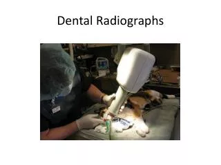
Interpreting Radiographs
Interpreting Radiographs. What to Look For. Dental caries Periapical pathology Calcified pulps Changes in alveolar bone pattern Alveolar bone height. What to Look For (Cont'd). Etiologic agents that promote dental disease Overhanging margins on restorations Calculus Open contacts.

Interpreting Radiographs
E N D
Presentation Transcript
What to Look For • Dental caries • Periapical pathology • Calcified pulps • Changes in alveolar bone pattern • Alveolar bone height
What to Look For (Cont'd) • Etiologic agents that promote dental disease • Overhanging margins on restorations • Calculus • Open contacts
Root caries may be confused with cervical burnout (see below). Cervical Burnout Cross-section (red line at right)
Cervical burnout Radiolucency seen above left (arrow) disappears on periapical film of same tooth (above right).
Anterior Cervical Burnout bone level cervical burnout area
Cervical burnout in the anterior region due to gap between enamel (red arrows) and alveolar bone over root (blue arrows).
Normal Alveolar Bone • Height 1.5 to 2 mm below CEJ of adjacent teeth • Bone forming alveolar crest should be smooth and intact • Slight radiolucent space adjacent to the root surface • Visible distinct crestal lamina dura
CEJ 1-1.5 mm
Alveolar crests more pointed anteriorly
Periodontal Disease Vertical Bitewings or paralleling PA’s best for diagnosis. Higher kVp recommended (long scale, low contrast). Compare images from different visits (using same technique).
Limitation of Radiographs Two-dimensional film with overlapping bony walls, superimposed roots Clinical picture more advanced Relationship of hard to soft tissues not evident
Periodontitis Involvement: Localized Generalized
Horizontal bone loss: Parallel to line drawn between adjacent CEJ’s Vertical (Angular) bone loss: More bone destruction on interproximal aspect of one tooth than on the adjacent tooth
Contributing Factors • Occlusal trauma • Open contacts • Overhangs, poor contours • Calculus • Post-extraction defects • Systemic involvement (diabetes, • blood disorders, hormonal • changes, stress, AIDS)
Gingivitis No bone loss No radiographic signs
Recognizing Early Alveolar Bone Loss • Increasing area of radiolucency where the alveolar crest and PDL meet • Widening periodontal ligament space • Loss of integrity of crestal lamina dura
Moderate adult periodontitis (red arrows point to calculus)
Restorative Materials Radiopaque: Structures with higher object density, such as amalgam, gold, silver points, pins, gutta percha, porcelain. Radiolucent: Structures with lower object density, such as older composites and bonding agents.
porcelain crowns
crown crown amalgam cast post gutta percha silver points