Download
1 / 25
360 likes | 726 Views
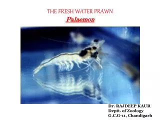
THE FRESH WATER PRAWN Palaemon. Dr. RAJDEEP KAUR Deptt . of Zoology G.C.G-11, Chandigarh. CONTENTS. Natural History Habitat Habits External characters Exoskeleton Morphology and Physiology Body divisions Cephalothorax Abdomen

E N D
THE FRESH WATER PRAWNPalaemon Dr. RAJDEEP KAUR Deptt. of Zoology G.C.G-11, Chandigarh
CONTENTS Natural History Habitat Habits External characters Exoskeleton Morphology and Physiology Body divisions Cephalothorax Abdomen Digestive System Respiratory System Circulatory System Excretory System Nervous system Reproductive system
NATURAL HISTORY HABITAT • Inhabits fresh water streams, rivers, lakes and ponds in Central and South India. HABITS • Nocturnal. • Omnivorous ( feed on small weeds, insects, algae). • Respiration through gills. • Sexual mode of Reproduction. • Dual mode of locomotion: a. Walking b. Swimming
Entire body of prawn is covered by a calcareous shell or exoskeleton. The colour of the shell comes by the deposition of lime salts and sclerotin. It comprises of several hardened plates, called sclertieswhich are connected with each other by a thin soft uncalcified cuticle or the arthrodial membrane, making the movement feasible. (a)Cephalothoracicsclerites: • All the cephalothoracicscleritesform a single large and continuous dorsal shield which extends over the head as a laterally compressed, serrated process called rostrum. • At the base of rostrum a pair of orbital notch is found that lodged a stalked, movable, compound eye. Just behind the orbital notch there are two outgrowths, the anterior antennal spine and the posterior hepatic spine. • The posterior region of dorsal sheild is called carapace. On either side of thorax it hangs down freely as branchiostegite or gill cover. (b)Abdominal sclerites: • Each abdominal segment is covered by separate sclerites which are joined with each other by arthrodial membrane . Each sclerite is composed dorsally by tergum, ventrally by narrow bar like plate sternum and the two flap like lateral plates pleura. • Abdominal appendages are connected with pleuron by a small plate epimeron. Tergum and pleura of adjacent segments slightly overlap each other and this arrangement is called imbricate arrangement. • Two adjacent abdominal segments articulate with each other by means of a pair of hinge joints. However, the hinge joints are lacking between the third and fourth segment.
A summary of the segments and the functions of each appendage
BODY WALL • Cuticle a. Epicuticle (thin outer layer). b. Procuticle(thick inner layer). • Epidermis Single layer of columnar cells with gland cells here and there. • Basement Membrane Single layer of flattened cells. The body wall bears setae and spines.s FUNCTIONS • Maintain body form. • Check loss of water. • Protect delicate internal organs fron injury, harmful chemicals and entry of microorganisms. • Helps in respiration. • Outgrowths forms sensory, defensive and feeding apparatus.
DIGESTIVE SYSTEM ALIMENTARY CANAL The alimentary canal is a long tube, which extends from the mouth to the anus. It is divisible into three regions. Stomodaeum: Anterior region lined with chitin and consists of mouth, buccal cavity, oesophagus and stomach.Themouth lies on the ventral side of the cephalothorax, between the mandibles. It leads into a short buccalcavity which are connected by oesophagus.Stomach consists of an anterior larger cardiac and the posterior smaller pyloric stomach. The inner dorso-lateral folds of this region bear stout denticleswhich form the gastric armature or gastric mill for grinding the food. Mesenteron:The pyloric stomach opens into the mesenteron which is not lined with chitin. The chitinous lining of the pyloric stomach forms one median dorsal, one median ventral and two lateral lippets or valvulae. Proctodaeum: The proctodaeum is the posterior region of the alimentary canal. It is lined with chitin. It consists of the hindgut (or rectum) and the anus. The anus lies at the base of the telson ventrally.
DIGESTIVE GLAND The digestive gland is large, orange red structure and is known as the hepato-pancreas or liver which lies in the cephalothoracicregion. It consists of two separate lobes called hepatic caeca. It. The digestive secretions of this gland reach the mesenteron by a pair of openings. PHYSIOLOGY OF DIGESTION Ingestion:Intake of food is aided by cephalothorax appendages. The mandibles cut the food into small pieces. The maxillae and maxillipeds aid in swallowing the food. In the buccal cavity food is masticated and then passes into the cardiac stomach through peristaltic movements of oesophagus. Digestion:Digestive juice secreted by hepatopancreas met with masticated food in stomach. It contains amylolytic, lipolytic and proteolytic enzymes that digest the starches, fats and proteins respectively. Absorption: Absorption takes place in the hepatopancreas and the intestine. Egestion: The hindgut forms faecal pellets, which are then passed out through the muscular anus.
RESPIRATORY SYSTEM Palaemontakes up oxygen dissolved in sea-water. Its respiratory organs are inner lining of branchiostegites, epipodites (mastigobranchiae) and branchiae (gills). • Branchiostegites: The ventral extension of the carapace on either side of cephalothorax is known as branchiostegite. The space between the bodywall and branchiostegite is called gill chamber. The inner lining of branchiostegite is highly vascular. It is bathed in water where exchange of respiratory gases takes place. • Epipodites:There are six pairs of epipodites. They are the outgrowth of coxae of the thoracic appendages, three pairs of maxillipeds and three pairs of chelate walking legs. They lie in the branchial chamber. They are bathed in water and richly supplied with blood. Here, exchange respiratory gases between water and blood takes place. • Branchiae or Gills: Branchiae(gills) are the feather like (plumose ) outgrowth of the lateral wall of the thorax and thoracic appendages. Each gill has a stem. Three longitudinal blood channels run through the stem. They are two lateral channels and one median channel. The two lateral channels are connected by many transverse channels. Many lateral flat gill plates arise from the stem. Marginal channels from the lateral channels penetrate into the gill plate and open into the median channel, The stem and gill plate are covered externally a thin layer of chitin. A single layer of epithelial cells lies beneath it. Epithelial layer encloses connective tissue and blood channels. This kind of gill is known as dendrobranchia.
MECHANISM OF RESPIRATION The scaphognathite of each maxilla lies anteriorly inside the gill chamber. By its constant vibrating movements it bales out water from the anterior open end of gill chamber. Action of scaphognathite is supplemented by the exopodites of maxillpedes. Fresh water enters the gill chamber from behind in the form of a current. This current of water flows over the lining of branchiostegite gills & epipodites which are richly supplied with blood so that exchange of gases takes place. The extremely delicate thin gill plates act as excellent permeable membrane for the passage of gases to & fro through diffusion. Oxygen dissolved in water is taken in by blood & CO2from blood diffuses out in the water.
CIRCULATORY SYSTEM Circulatory system consists of heart, arteries, pericardial membrane, pericardial sinus, haemocoel, blood channels and blood or haemolymph. HEART:Theheart is a triangular chamber. It lies in the pericardial space. It is provided with paired openings called ostia. ARTERIES: Arteries are the main tubes which arise from the anterior and posterior regions of the heart. These are opthalmic artery, antennary arteries, hepatic arteries, sternointestinal artery
PERICARDIAL SINUS: It is a large space in the dorsal part of thorax and contains the heart in it. HAEMOCOEL: The spaces between the visceral organs form the haemocoel. It contains blood or haemolymph. From the haemocoel blood goes to the gills through the blood channels. From the gills blood goes to the heart through blood channels. The blood contains plasma, haemocytes or blood cells and the respiratory pigment haemocyanin. CIRCULATION OF HAEMOLYMPH Haemolymph in the pericardial sinus enters the heart through the ostia. From the heart it reaches reaches the haemocoel through the arteries. Then it is collected by the afferent blood channels and returned to the pericardial sinus through the gills and efferent blood channels.
NERVOUS SYSTEM BRAIN. A. Dorsal view. B. Ventral view NERVOUS SYSTEM
CENTRAL NERVOUS SYSTEM It consists of supraoesophageal ganglion or brain, circumoesophagealconnectives and nerve cord. • Supraoesophageal Ganglia: It lies under the base of the brain. The brain is a composite structure represents fused pair of ganglia. • Circumoesophageal Connectives: They arise from lateral parts ogbrainand pass backward and downward round the oesophagus to meet the ventral nerve cord. • Nerve cord: Nerve cord is lies in midventral line of the body. It arises from the sub oesophageal ganglion and runs upto the end of the abdomen. It bears 17 pairs of ganglia, one belonging to each postoral segment. The anterior 11 pairs belong to cephalothorax. The posterior 6 pairs belong to abdomen.
2. PERIPHERAL NERVOUS SYSTEM It consists of paired nerves that arise from various parts of central nervous system to innervate the body parts. These are • Optic nerves • Ophthalmic nerves • Antennulary nerves • Antennary nerves • Tegumental nerves • Mandibular nervess • Maxillary nerves • Pedal nerves 3. AUTONOMIC NERVOUS SYSTEM It consist of nerve arise fromhind end of brain and bears two small visceral ganglia. The anterior visceral ganglion is connected with the commissural ganglia borne by the circumoesophageal connectives by transverse connectives. The posterior visceral ganglia gives off two pairs of nerves to the muscles of wall of oesophagus and stomach.
SENSE ORGANS • Compound eyes: There are two compound eyes/ one on either side of the base of the rostrum. They are at the ends of movable stalks. Each eye is made up of many optical units called simple eyes or ommatidia. Each ommatidium consists of an outermost layer called cornea. It is formed by the transparent cuticle. Externally this layer is hexagonaland called a facet. Two types of images are formed.Theyare mosaic (apposition) and super-imposed (superposition) images. • Statocysts:There is a pair of statocysts. They are organs of orientation and equilibrium. Each statocyst lies at the basal segment of the antennule. It is a sac- like structure filled with sand particles which function as statolith. The sand particles are surrounded by elongated delicate receptor setae. This sts up a nerve impulse ehich is conveyed to brain by nerve. • Tactile organs and setae : Antennae are the important tactile sense organs. Many sensory setae are located over the body surface especially the appendages. • Olfactory setae: They are present on the middle small feeler of each antennule.
EXCRETORY SYSTEM The excretory system consists of • Antennal glands:There is a pair ofantennal or green glands. Each lies enclosed in the proximal segment (coxa) of the antenna. Its parts are an end sac, a coiled tube and a bladder. • Renal sac:The renal sac is large, blind. It cover the cardiac stomach and reach the gonads. Anteriorly, it communicates with bladder. The tubular part is glandular and the bladder is thin walled. The bladder opens to the exterior by the excretory pore.
PHYSIOLOGY OF EXECRETION • EXCRETION: The green glands have good blood supply from antennary arteries, by ultrafilteration, water and dissolved substances passing from surrounding blood into endsacs. The filterate, called primary urine which passes into labyrinths. Here, selective resorption takes place where useful material pass into blood and remaining fluid is called final urine which flows into bladder. The urine is passed out through ureters and renal apertures and is wafted away in the currents set up by scaphognathites. • OSMORUGULATION:Prawn blood is hypertonic to surrounding water. Therefore water diffuses into blood through highly permeable gills, that is why prawn passes out a large quantity of hypotonic urine to regulate internal fluid volume.
REPRODUCTIVE SYSTEM • Male Reproductive System: • Testes: There are two testis situated over posterior half of hepatopancreas and beneath heart. They extend upto renal sac in abdomen. Each testis is made up of large number of lobules held together by connective tissue. They are tubular and united in front. Many caecal diverticula arise from each testis. • Vasa deferentia:From the posterior region of each testis arises a tubular structure called vas deferens. It has a narrow anterior region, a swollen and convoluted middle region and a narrow posterior region. • Vesiculaeseminales: These are club shaped posterior ends of the vasa deferentia. They store spermatozoa packed in spermatophores. • Male genital openings: The openings of the vesiculaseminales to the exterior are called the male genital openings.
Female reproductive system: • Ovaries: There are two ovaries, one on either side of the middle line and they occupy the whole length of the thorax and abdomen. The right and left ovaries are united posteriorly and free anteriorly. Many distinct diverticula arise from the anterior region of the ovaries. • Oviducts: These are selender curved tubes with wide proximal ends. From the middle region of each ovary arises an oviduct. • Female genital openings: The two oviducts open to the exterior by the female genital openings.
LIFE HISTORY The female releases the eggs in the water. Nauplius larva emerges from the egg. It passes the metanaupleusprotozoea, mysis, postlarval, juvenile stages and becomes the adult.
ECONOMIC IMPORTANCE Fresh, and nutritious seafood is always the spirit for various delicious dishes. Either serving as live, fresh, boiled, steamed, chilled, fried, or baked, seafood is always the popular dishes in many restaurants. Prawns are the major high priced seafood. The function of the marine creatures is not restricted to as seafood only, but many underlying uses are still remained to be discovered. In old ages, some have been used as a horn for recreational use. Some of the colorful species are collected as ornaments in containers. Some are used for jewelry, buttons and inlays. The shell of many molluscs can be sold for the manufacture of lime. Moreover, many crabs can serve as feed for fish cultures, for turtles and domestic birds and other animals. On duck farms, food mixed with crab powder not only stimulates growth and the fattening of ducks, but may also raise their spawning rate. Furthermore, dried seafood are on sale and widely used by Asian people as soup ingredients. Because they believe that seafood have potent medical contribution to good health. In conclusion, it is undeniable that marine invertebrates are important, both in its delicacy as seafood and its tremendous economic value.