Download
1 / 24
270 likes | 496 Views
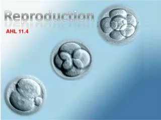
11.4 Reproduction Higher. A mature Sperm. You need to know the top diagram. The bottom ones are included for interest only!.

E N D
A mature Sperm You need to know the top diagram. The bottom ones are included for interest only! http://images.google.com.ph/imgres?imgurl=http://upload.wikimedia.org/wikipedia/commons/thumb/0/03/Complete_diagram_of_a_human_spermatozoa.svg/180px-Complete_diagram_of_a_human_spermatozoa.svg.png&imgrefurl=http://en.wikibooks.org/wiki/Human_Physiology/The_male_reproductive_system&usg=__zSltk3LvardHYMqGrfqBWtXJjxo=&h=176&w=180&sz=18&hl=en&start=3&tbnid=U7gKJCUpYC4dmM:&tbnh=99&tbnw=101&prev=/images%3Fq%3Dspermatazoa%2Bdiagram%26gbv%3D2%26hl%3Den http://www.bmu.unimelb.edu.au/resources/illustrations/images/sperm_thumb.gif
A mature ovum Nucleus Nucleolus Membrane with cortical granules Cytoplasm containing lipid granules www.gettyimages.com/detail/BC8592-001/Stone Zona pellucida http://etc.usf.edu/clipart/50700/50718/50718_ovum_lg.gif
Fertilisation • 2 reactions occur in fertilization. The acrosome reaction and the cortical reaction As the sperm approaches the zona pellucida of the egg, which is necessary for initiating the acrosome reaction, the membrane surrounding the acrosome fuses with the plasma membrane of the sperm, exposing the contents of the acrosome. The contents include surface antigens and numerous enzymes which are responsible for breaking through the egg's tough coating and allowing fertilization to occur. http://images.google.com.ph/imgres?imgurl=http://upload.wikimedia.org/wikipedia/commons/thumb/2/26/Acrosome_reaction_diagram.svg/440px-Acrosome_reaction_diagram.svg.png&imgrefurl=http://en.wikibooks.org/wiki/Human_Physiology/The_male_reproductive_system&usg=__X1gzuaYckkk_8BRocULr7-jhhEY=&h=310&w=440&sz=72&hl=en&start=2&tbnid=wvYtqlT1PP6GVM:&tbnh=89&tbnw=127&prev=/images%3Fq%3Dspermatazoa%2Bdiagram%26gbv%3D2%26hl%3Den
The cortical reaction, also known as the zona reaction, occurs when a sperm cell unites with the egg's plasma membrane, altering the zona pellucida which prevents other sperm from binding to and entering the egg. • The cortical reaction is exocytosis of the egg's cortical granules. Cortical granules are secretory vesicles that reside just below the egg's plasma membrane. When the fertilizing sperm contacts the egg plasma membrane, it causes calcium to be released from storage sites in the egg, raising the intracellular free calcium concentration. This triggers fusion of the cortical granule membranes with the egg plasma membrane, liberating the contents of the granules to the extracellular space. Fusion begins near the site of sperm contact, and then as the wave of calcium release sweeps around the egg, a wave of cortical granule fusion results. The contents of the granules vary with the species, and are not fully understood. http://en.wikipedia.org/wiki/Cortical_reaction
http://faculty.southwest.tn.edu/rburkett/A&P2_r7.jpg http://www.kumc.edu/instruction/medicine/anatomy/histoweb/male/male04.htm
The small cells at the base of the seminiferous epithelium are spermatogonia. • The ones with the largest are nuclei are primary spermatocytes. • There are spermatids (round & elongating) about to be released into the lumen.
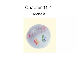
Spermatogenesis • Spermatogonia push their way through the tight junctions between adjacent Sertoli cells and become primary spermatocytes. B. In the first meiotic division each of the 46 chromosomes doubles up and then forms a pair with another doubled up chromosome during which time there is exchange of genetic material (crossing over). The cell divides and each daughter cell contains one of the pairs of chromosomes. In the second meiotic division each double chromosome simply separates producing genetically identical secondary spermatocytes.
The first week of pregnancy. Use this diagram for the timings. The labels are beyond your IB requirements. http://stemcells.nih.gov/StaticResources/info/scireport/images/figurea2.jpg
The embryo goes through its cleavage stages while it is migrating along the fallopian tube. As a blastocyst it reaches the uterine cavity at the end of the 5th day. There, the hatching and the embedding into the endometrium occur on the 6th day. http://mcb.berkeley.edu/courses/mcb135e/Difficult%20Slides/Fertilization%201.jpg
The morula, a collection of around 30 cells (blastomere), is created at about 96 hours. Because these cells arise only through the cleavage of the zygote and all are found inside the pellucid zone, which cannot expand, no growth is seen. Every new cell is thus only half as large as the cell from which it derives. The name of this stage comes from its resemblance to a mulberry, since it really looks like a collection of spherical cells. http://www.embryology.ch/anglais/evorimplantation/furchung01.html
A blastocyst The blastocyst comes into being through compaction of the cells and the accumulation of intercellular fluid, leading to the formation of the blastocyst cavity. At this point, the embryoblast that lies inside (hump on the left side) consists of roughly 12 cells. At the same time, the enveloping trophoblast, made of a single cellular layer, contains around a hundred cells. Hatching Around the end of the fifth day the embryo frees itself from the enveloping pellucid zone. Through a series of expansion-contraction cycles the embryo bursts the covering. This is supported by enzymes that dissolve the pellucid zone. The rhythmic expansions and contractions result in the embryo bulging out of and emerging from the rigid envelope. This "first birth" is called hatching.
Implantation Having ‘hatched’ the blastocyst nows burrows into the endometrium. The inner mass (embryoblast) starts to develop into the embryo. The trophoblast develops into the amniotic sac and placenta. http://mcb.berkeley.edu/courses/mcb135e/Difficult%20Slides/Implantation%203.jpg
Early embryo After 4 weeks Implantation site. The basic structure of the placenta has been formed with maternal blood being delivered to the forming placenta via spiral arteries while being drained away via uterine veins. Like the roots of a tree, the developing chorionic villi remain immersed in a space filled with the nutrient rich maternal blood. http://www.med.yale.edu/obgyn/kliman/placenta/articles/EOR_Placenta/Trophtoplacenta.html
The amnion secretes amniotic fluid that protects the baby. It acts as a physical shock absorber and due to its high water content (and the high Spec.Heat.Cap. of the water) as a thermal buffer. http://www.colorado.edu/intphys/Class/IPHY3430-200/image/26-19.jpg The placenta allows exchange of materials between the mother and embryo without allowing the blood to mix. It also produces Progesterone and oestrogen.
The hormonal control of birth. Oxytocin causes the muscles of the uterus to contract. And prepare the mammary glands to secrete milk. http://www.gynaeonline.com/images/pituitary_gland.gif Stretch receptors in the uterus send a signal to the pituitary gland to release oxytocin into the blood stream. http://chuvachienes.com/wp-content/uploads/2009/09/40-weeks-pregnant-internal.gif This is called Positive feedback.
Birth http://www.healthsquare.com/fgwh/wh1c2601.jpg
Now watch these videos…. • http://www.pbs.org/wgbh/nova/miracle/program.html