Understanding Testis Tissue: Cell Types & Spermatogenesis Process
320 likes | 446 Views
This document provides a detailed annotation of a light micrograph of testis tissue, highlighting the location and function of various cell types such as Leydig cells, germinal epithelium cells, Sertoli cells, and developing spermatozoa. It explains the role of interstitial cells in testosterone secretion, the processes of spermatogenesis including cell division and differentiation, and the influence of hormones like LH and FSH. Additionally, it outlines the anatomy of the testis and provides insights into cellular development relevant to male reproductive biology.

Understanding Testis Tissue: Cell Types & Spermatogenesis Process
E N D
Presentation Transcript
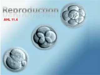
Reproduction 11.4 IB STANDARDS PART II Bianca arguilla Brea alotaya Natasha guenther
11.4.1: Annotate a light micrograph of testis tissue to show the location and function of interstitial cells (leydig cells), germinal epithelium cells, developing cells, developing spermatozoa, and Sertoli cells • Parts to know on the testis tissue: • wall of seminiferous tubule • Fluid inside seminiferous tubule • Blood vessel • Interstitial cells (Leydig cells) secrete testosterone
11.4.1: Annotate a light micrograph of testis tissue to show the location and function of interstitial cells (leydig cells), germinal epithelium cells, developing cells, developing spermatozoa, and Sertoli cells • Interstitial Cells (Leydig cells): adjacent to the seminiferous tubules in the testicle. They can secrete testosterone and are often closely related to nerves. Leydig cells have round vesicular nuclei and an eosinophilic cytoplasm • Leydig cells release a class of hormones called androgens. They secrete testosterone, androstenedione and dehydroepiandrosterone (DHEA), when stimulated by the pituitary hormone LH. LH increases cholesterol desmolase activity, leading to testosterone synthesis secretion by Leydig cells. • FSH increases the response of Leydig cells to LH by increasing the number of LH receptors expressed on Leydig cells. • Germinal epithelium (male): innermost layer of the testicle • cellular covering of internal and external surfaces of the body, including the lining of vessels and other small cavities. It consists of cells joined by small amounts of cementing substances
11.4.1: Annotate a light micrograph of testis tissue to show the location and function of interstitial cells (leydig cells), germinal epithelium cells, developing cells, developing spermatozoa, and Sertoli cells • Developing cells: are able to revert back into an embryonic-like stem cell state, which could then be driven into chosen cell types; can continue to produce sperm • In science today, testis tissues have been found to be “stem-like” cells that are capable of being harvested and used in the future • developing spermatozoa: aka sperm • Spermatogenesis is the production of spermatozoa and occurs in the narrow tubes called seminiferous tubules in the testes • Sertoli cells: a 'nurse' cell of the testes which is part of a seminiferous tubules; nurtures developing sperm cells through spermatogenesis • It is activated by FSH and has FSH receptors on its membranes.
11.4.2: OUTLINE THE PROCESSES INVOLVED IN SPERMATOGENESIS WITHIN THE TESTIS, INCLUDING MITOSIS, CELL GROWTH, THE TWO DIVISIONS OF MEIOSIS, AND CELL DEFFERENTIATION
11.4.2: OUTLINE THE PROCESSES INVOLVED IN SPERMATOGENESIS WITHIN THE TESTIS, INCLUDING MITOSIS, CELL GROWTH, THE TWO DIVISIONS OF MEIOSIS, AND CELL DEFFERENTIATION • 6 Stages total: • 1. An outer layer called the germinal epithelium cells [2n] divide endlessly by MITOSIS to produce more diploid cells • 2. Diploid cells GROW larger and are then called primary spematocytes[2n] • 3. Each primary spermatocyte carries out the FIRST DIVISION OF MEIOSIS to produce two secondary spermatocytes [n] • 4. Each secondary spermtocyte carries out the SECOND DIVISION OF MEIOSIS to produce two spermatids [n] • 5. Spermatids become associated with nurse cells, sertoli cells, which help the spermatids to develop into spermatozoa[n]. This is CELL DIFFERENTIATION. • 6. Sperm detach from sertoli cells and eventually are carried out of the testis by the fluid in the centre of the seminiferous tubule.
11.4.2: OUTLINE THE PROCESSES INVOLVED IN SPERMATOGENESIS WITHIN THE TESTIS, INCLUDING MITOSIS, CELL GROWTH, THE TWO DIVISIONS OF MEIOSIS, AND CELL DEFFERENTIATION
11.4.3: STATE THE ROLE OF LH, TESTOSTERONE, AND FSH IN SPERMATOGENESIS
11.4.4: ANNOTATE A DIAGRAM OF THE OVARY TO SHOW THE LOCATION AND FUNCTION OF GERMINAL EPITHELIUM, PRIMARY FOLLICLES, MATURE FOLLICE, AND SECONDARY OOCYTE • Parts to know on the ovary: • Region where blood vessels enter and leave • Outer layer of germinal epithelium cells • Cortex [containing primary follicles] • Secondary oocyte inside a mature follicle • Medulla [containing blood vessels]
11.4.4: ANNOTATE A DIAGRAM OF THE OVARY TO SHOW THE LOCATION AND FUNCTION OF GERMINAL EPITHELIUM, PRIMARY FOLLICLES, MATURE FOLLICE, AND SECONDARY OOCYTE • Germinal Epithelium: surface of the ovary covered by a layer of simple cuboidal cells • These cells are derived from the mesoderm during embryonic development and are closely related to the mesothelium of the peritoneum. The germinal epithelium gives the ovary a dull gray color as compared with the shining smoothness of the peritoneum; and the transition between the mesothelium of the peritoneum and the columnar cells which cover the ovary is usually marked by a line around the anterior border of the ovary. • The germinal epithelium gives rise to primary follicles • Primary Follicles: Located inside the cortex; develop receptors to FSH at this time, but they are gonadotropin-independent up until the latter stages
11.4.4: ANNOTATE A DIAGRAM OF THE OVARY TO SHOW THE LOCATION AND FUNCTION OF GERMINAL EPITHELIUM, PRIMARY FOLLICLES, MATURE FOLLICLE, AND SECONDARY OOCYTE • Mature Follicle: contains secondary oocyte; bursts open and releases the egg with increased levels of LH, starting ovulation • Secondary Oocyte: An oocyte in which the first meiotic division is completed. The second meiotic division usually stops short of completion unless fertilization occurs; b/w first and second maturation development
11.4.5: OUTLINE THE PROCESSES INVLOVED IN OOGENESIS WITHIN THE OVARY, INCLUDING MITOSIS, CELL GROWTH, THE TWO DIVISIONS OF MEIOSIS, THE UNEQUAL DIVISION OF CYTOPLASM, AND THE DEGENERATION OF POLAR BODY
11.4.5: OUTLINE THE PROCESSES INVLOVED IN OOGENESIS WITHIN THE OVARY, INCLUDING MITOSIS, CELL GROWTH, THE TWO DIVISIONS OF MEIOSIS, THE UNEQUAL DIVISION OF CYTOPLASM, AND THE DEGENERATION OF POLAR BODY • 8 STEPS: • 1. In the ovaries of a female fetus, germinal epithelium cells [2n] divide by MITOSIS to form more diploid cells [2n] • 2. Diploid cells GROW into larger cells called primary oocytes [2n] • 3. Primary oocytes start the FIRST DIVISION OF MEIOSIS but stop during the prophase I. The primary oocyte and a single layer of follicle cells around forma primary follicle • 4. When a baby girl is born the ovaries contain about 400,000 primary follicles • 5. Every menstrual cycle a few primary follicles start to develop. The primary oocyte completes the first division of meiosis, forming two haploid nuclei. The cytoplasm of the primary oocyte is DIVIDED UNEQUALLY forming a large secondary oocyte [n] and a small polar cell [n] • 6. The secondary oocyte starts the SECOND DIVISION OF MEIOSIS but stops in prophase II. The follicle cells meanwhile are proliferating and follicular fluid is forming. • 7. When the mature follicle bursts, at the time of ovulation, the egg that is released is actually still a secondary oocyte. • 8. After fertilization the secondary oocyte completes the second division of meiosis to form an ovum, [with a sperm nucleus already inside it] and a second polar cell or body. The first and second polar bodies do not develop and eventually DEGENERATE.
11.4.5: OUTLINE THE PROCESSES INVLOVED IN OOGENESIS WITHIN THE OVARY, INCLUDING MITOSIS, CELL GROWTH, THE TWO DIVISIONS OF MEIOSIS, THE UNEQUAL DIVISION OF CYTOPLASM, AND THE DEGENERATION OF POLAR BODY
11.4.6: DRAW AND LABEL A DIAGRAM OF A MATURE SPERM AND EGG FEATURES TO LABEL: HEAD, ACROSOME, HAPLOID NUCLEUS, MID-PIECE, TAIL, PROTEIN FIBERS TO STRENGTHEN TAIL, MICROTUBULES IN A 9+2 ARRANGEMENT, HELICAL MITOCHONDRIA, CENTRIOLE, PLASMA MEMBRANE
11.4.6: DRAW AND LABEL A DIAGRAM OF A MATURE SPERM AND EGG FEATURES TO LABEL: TWO CENTRIOLES, FIRST POLAR CELL, PLASMA MEMBRANE, LAYER OF FOLLICLE CELLS [CORONA RADIATA], LAYER OF GEL COMPASED OF GLYCOPROTEINS [ZONA PELLLUCIDA], CORTICAL GRANULES, CYTOPLASM/YOLK CONTAINING DROPLETS OF FAT, HAPLOID NUCLEUS
11.4.7: OUTLIHNE THE ROLE OF THE EPiDIDYMIS, SEMINAL VESICLE, AND PROSTATE GLAND IN THE PRODUCTION OF SEMEN • The three main structures: Epididymis, Seminal vesicles, and Prostate gland • When sperm first arrive in the epididymis from the testes, they are unable to swim • While in epididymis, sperm matures and learn to swim • The prostate gland and two seminal vesicles produce and store fluids that are later expelled during ejaculation • The fluid them mixes with them sperm and increased the volume of the ejaculate • The fluid from the seminal vesicle contains nutrients, like fructose, for the sperm and a muscus to protect it in the vagina • The fluid from the prostate gland has mineral ions and protects the sperm from the vagina’s acidic conditions due to its alkalinity.
COMPARE THE PROCESSES OF SPERMATOGENESIS AND OOGENESIS, INCLUDING THE NUMBER OF GAMETES AND THE TIMING OF THE FORMATION AND RELEASE OF GAMETES • Similarities: • Both start with the proliferation of cells by mitosis • Both involve the cell growth before mitosis • Both involve the two divisions of meiosis • Differences:
11.4.9: DESCRIBE THE PROCESS OF FERTILIZATION, INCLUDING THE ACROSOME REACTION, PENETRATION OF THE EGG MEMBRANE BY A SPERM AND THE CORTICLE RXN
11.4.9: DESCRIBE THE PROCESS OF FERTILIZATION, INCLUDING THE ACROSOME REACTION, PENETRATION OF THE EGG MEMBRANE BY A SPERM AND THE CORTICLE RXN • 6 steps total: • 1. Arrival of sperm: Sperm attracted by a chemical signal and swim up the oviducts to reach the egg. Fertilization is only successful if many sperm reach the egg {sperm tries to push through the layers of follicle cells around the egg} • 2. Binding: The 1st sperm to break through the layers of follicle cells binds to the zonapellucida. This triggers the acrosome reaction. • 3. Acrosome Reaction: The contents of the acrosome are released by the separation of the acrosomal cap from the sperm. Proteases from the acrosome digest a route for the sperm through the zonapellucida, allowing the sperm to reach the plasma membrane of the egg
11.4.9: DESCRIBE THE PROCESS OF FERTILIZATION, INCLUDING THE ACROSOME REACTION, PENETRATION OF THE EGG MEMBRANE BY A SPERM AND THE CORTICLE RXN • Fertilization continued • 4. Fusion: The plasma membrane of the sperm and egg fuse and the sperm nucleus enters the egg and joins the egg nucleus. Fusion causes the cortical reaction. • 5. Cortical Reaction: Small vesicles called cortical granules move to the plasma membrane of the egg and fuse with it, releasing their contents by exocytosis. Enzymes from the cortical granules cause the cross linking glycoproteins in the zonapellucida, making it hard and preventing the entry of anymore sperm • 6. Mitosis: The nuclei from the sperm and the egg do not fuse together. Instead, both nuclei carry out mitosis using the same centrioles and spindle of microtubules. A 2-celled embryo is produced
11.4.10: OUTLINE THE ROLE OF HCG IN EARLY PREGNANCY • HCG: human chronic gonadotrophin; prevents degeneration of the corpus luteum. • Estrogen and progesterone are needed throughout pregnancy to stimulate the development of the uterus lining.During the first few days after ovulation, the corpus luteum secretes these hormones whether or not there has been fertilization. After implanting in the uterus wall, the embryo starts to secrete HCG. • HCG stimulates the corpus luteum to grow and to continue secretion of estrogen and progesterone. This is essential to allow the pregnancy to continue. • By the middle of the pregnancy, the corpus luteum starts to degenerate, but by then the cells in the placenta are secreting estrogen and progesterone and these cells secrete increasing amounts until the end of the pregnancy.
11.4.11: OUTLINE THE EARLY EMBRYO DEVELOMPENT UP TO THE IMPLANTATION OF THE BLASTOCYST • Fertilization: Fusion of the egg and the sperm • During copulation, or sexual intercourse, semen is ejaculated into the vagina. Sperm swim through the cervix, up the uterus and into the oviducts. If there is an egg in the oviducts, the sperm can fuse with it to produce a zygote • Zygotes produced in the oviduct = new individual • Zygote starts to divide by mitosis and forms a 2-cell embryo, and then a 4-cell embryo and so on until a blastocyst, or a hollow ball of cells, is formed • During these stages embryo is transported down the oviducts to the uterus {at about 7 days old the embryo implants itself into the wall of the uterus to continue development}.
11.4.12: EXPLAIN HOW THE STRUCTURE AND FUNCTIONS OF THE PLACENTA, INCLUDING ITS HORMONAL ROLE IN SECRETION OF ESTROGEN AND PROGESTERONE, MAINTAIN PREGANCY • Placenta: a disc-shaped structure, 185 mm in diameter and 20 mm thick when fully grown. • PlacentileVilli: small projections that give a large surface area, 14 m^2, for gas exchange and exchange of other materials. Fetal blood flows through the capillaries in the villi • Inter-villous Spaces: Maternal blood flows through these spaces, brought by the uterine arteries and carried away by the uterine veins. • Myometrium: muscular wall of the uterus used during childbirth. • Oxygenated Fetal Blood: flows back to the fetus from the placenta along the umbilical vein • De-oxygenated Fetal Blood: flows from the fetus to the placenta along the umbilical arteries • Endometrium: the lining of the uterus which the placenta grows
11.4.13: STATE THAT THE FETUS IS SUPPORTED AND PROTECTED BY THE AMNIOTIC SAC AND AMNIOTIC FLUID • The fetus develops an amniotic sac containing amniotic fluid around it. • The fetus floats in and is supported by the amniotic fluid b/c the fluid acts as a shock absorber (such as everyday events, or accidents that impacts the mother’s abdomen)
11.4.14: STATE THAT MATERIALS ARE EXCHANGED BETWEEN THE MATERNAL AND FETAL BLOOD IN THE PLACENTA • Background Info • 8 weeks+, embryo starts to develop tissue, becoming a “fetus.” The placenta and umbilical also develop. • The placenta has many projections called placenta villi embedded in the uterine wall. • In the placenta, the blood of the fetus flows close to the blood of the mother in the uterus wall. • Materials are exchanged between the maternal and fetal blood • Ex: Oxygen passes from maternal to fetal blood and Carbon Dioxide passes from fetal to maternal blood
11.4.15: OUTLINE THE PROCESS OF BIRTH AND ITS HORMONAL CONTROL, INCLUDING THE CHANGES IN PROGESTERONE AND OXYTOCIN LEVELS AND POSITIVE FEEDBACK • Over 9 months of pregnancy, progesterone ensure that the uterus develops and sustains the growing fetus • Levels are increasingly high until end of pregnancy in which a rapid decrease of the hormone occurs. • This causes secretion of oxytocinwhich causes the muscles of the uterus wall to contract • The uterine contractions then become stronger and stronger due to increase of oxytocin, creating a positive feedback • John Jacob Jingle Heimer Schmitt, his name was my name, too!