Download

1 / 40
420 likes | 1.22k Views
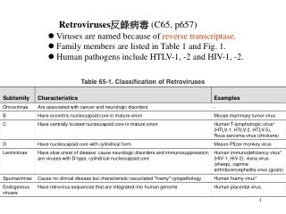
Reverse transcribing viruses other than retroviruses. Transposons. Class I - retrotransposons Very common in most organisms, especially plants Similar to retroviruses (e.g., HIV), but most particles don’t have envelopes and most are not infectious

E N D
Transposons • Class I - retrotransposons • Very common in most organisms, especially plants • Similar to retroviruses (e.g., HIV), but most particles don’t have envelopes and most are not infectious • Stress induced, are likely a major contributor to variation and evolution in all organisms • Two families classified with viruses - Metaviridae and Pseudoviridae, differ in gene order and relationships • Most do not contain env gene, but some do • Most have not been shown to be transmissible, some have • Class II - DNA mediated transposons • Well-known in bacteria, plants, and animals; more recently identified in fungi • Not classified with viruses
PstI PstI PstI PstI IR IR DR 1 DR 1 Transposition of Class II transposons (not classified with viruses; e.g., TN5 used for mutagenesis) Transposase Short ITRs; encode transposase 2
PstI PstI PstI PstI IR IR DR 1 DR 1 Transposition of Class II transposons Transposition is by cut-and-paste mechanism 2
PstI PstI PstI PstI Transposition of Class II transposons DR 1 DR 1 Circular DNA intermediate migrates to new position on same or different chromosome 2
PstI PstI PstI PstI Transposition of Class II transposons DR 1 DR 1 “Footprint” is evident because direct repeat sequences are left behind 2
PstI PstI PstI PstI Transposition of Class II transposons DR DR 2
PstI PstI PstI PstI Transposition of Class II transposons DR 1 DR 1 Different size restriction fragments are used to identify new insertions quickly, confirmed by sequence IR IR DR 2 DR 2
PstI PstI PstI PstI IR IR DR 1 DR 1 Transposition of Class II transposons When transposition occurs during meiosis, new and original copy may still be evident in progeny IR IR DR 2 DR 2
PstI PstI PstI PstI Transposition of Class I retrotransposons (classified with viruses) LTR LTR gag pol (RT) DR 1 DR 1 Have LTRs; encode gag (CP), pol (RT), RNaseH, protease, integrase; some have env 2
RNA PstI PstI PstI PstI Transposition of Class I retrotransposons LTR LTR gag pol (RT) DR 1 DR 1 Transposition is by transcription, reverse transcription, integration 2
RNA PstI PstI PstI PstI Transposition of Class I retrotransposons LTR LTR DR 1 DR 1 Transposition is by transcription, reverse transcription, integration DNA 2
RNA PstI PstI PstI PstI LTR LTR Transposition of Class I retrotransposons LTR LTR DR 1 DR 1 Transposition is by transcription, reverse transcription, integration DNA DR 2 DR 2
Properties of retrotransposons • Useful as genetic markers for population studies • May represent the majority of nuclear genome • They contribute to genetic variability and plasticity of organism • Transposition may be induced by environmental or developmental stresses • Knowing what induces transposition may allow one to predict behavior of organism in given conditions
Retrotransposon replication • Most of the general features of replication similar to retrovirus replication (see retrovirus lecture for details of replication) • For most, virion (without envelope) has been shown to be an essential part of replication cycle • Non-enveloped particle is not infectious under most circumstances - can’t leave cell
Retrotransposon properties • Two virus families: Metaviridae and Pseudoviridae differ in genome organizations and phylogenies • Genome sizes 4-13 kb • Some LTR have envelope gene (env), most do not • Infectivity has been demonstrated for those containing env protein • Horizontal transfer has been demonstrated for many transposons without env gene • No disease has been associated with infection and replication • Genetic disease or mutation may be associated with transposition • Transposition hot-spots may be found in host DNA
Non-LTR Retrotransposons Caulimoviridae Pseudoviridae Metaviridae Bacterial ms- associated DNA Mauriceville plasmid Hepadnaviridae GroupII Introns Retroviridae Phylogenetic relationships among reverse-transcribing elements Retrotransposons Retrotransposons from two families are not each other’s closest relatives In the figure, the length of the box indicates the relative diversity of reverse transcriptase genes within the family (longer is more diverse); the width of the box indicates the relative number of sequences used for the comparison (wider is more sequences). From Boeke et al., 2001 ICTV, 7th Report
Phylogenetic relationships among reverse-transcribing elements
Phylogenetic relationships among reverse-transcribing elements Color code: orange, DD site of RT; purple, RNase domain (RH); yellow, integrase domain (INT); blue, cysteine-histidine motif (C-H); green protease domain (PR); pink, envelope/movement protein domain (ENV/MP). 25From Hansen and Heslop-Harrison. 2004. Adv.Bot.Res.
Caulimoviridae • Isometric 50 nm T=7 particles or bacilliform particles; no envelope • Nicked dsDNA genomes ~ 8 kb • First plant virus shown to have DNA genome • Replication is by reverse transcription • Transcription is in nucleus; DNA replication in cytoplasm • Most do not integrate into host genome
Caulimoviridae • Most have narrow host ranges • Most are relatively unimportant as pathogens; exception is Rice tungro bacilliform virus, part of the most important rice virus complex • Most are transmitted by invertebrate vectors • Viruses do not replicate in vector; use virus-coded helper protein to aid transmission • Promoter elements commonly used in genetic engineering of plants • Caulimoviruses are not very versatile as plant gene expression vectors because of packaging constraints/instability
Visualization of Viral Particles • Electron micrographs of thin sections of caulimovirus-infected tissue showing 42-46 nm virus particles and inclusion bodies. (Photos courtesy of Dr. T.A. Chen)
Virus enters plant cell, capsid protein is removed • dsDNA enters nucleus; gaps closed; transcription to 35S and 19S RNAs • In the cytoplasm, the 19S RNA is translated to produce protein that forms inclusion bodies • Five ORFs are translated from 35S RNA by complex combination of strategies • Other copies of 35S RNA are reverse transcribed and packaged into virions • Particles exit cells through plasmodesmata or by aphids Caulimovirus Life Cycle Helper factor Inclusion Mature particle with DNA (Shaw, Fund. Virology, 3rd Ed., 1996)
Cauliflower mosaic virus genome structure • Seven ORFs on CaMV genome • Translation of seven proteins from two transcripts • ORF 2 is the only dispensable ORF • ORFs 6 and 7 are involved in translation regulation • Packaged genomic DNA has discontinuities on both strands • Replication is from tRNAmet primer Translation regulator Movement protein Helper component Inclusion, transactivator Replication factor Coat protein Reverse transcriptase
Genome Organization of Caulimoviridae Petuvirus I II PVCV Caulimoviruses VII I II III IV V VI CaMV FMV CERV Soymoviruses VII I a b c IV V VI SbCMV BRRV Cavemoviruses I II III IV V CsVMV Tungroviruses I II III IV RTBV Badnaviruses I II III ComYMV
Hepadnaviruses Hepatitis B
Hepatitis Viruses & Liver Disease The Liver is a target for six viruses that cause acute hepatitis. These are Hepatitis Virus A, a picornavirus passed in a fecal-oral pattern, the pararetrovirus Hepatitis B, that is transmitted via blood products, IV drug use, from mother to child & by unprotected sex, Hepatitis C, a flavivirus transmitted via IV drug use & earlier by blood products, Hepatitis Delta, a viroid-like satellite associated with HBV and Hepatitis E, a calicivirus Fig. 24.01 passed via feces contaminated water. Hepatitis G is a relatively rare flavivirus transmitted via blood and possibly during sex.
Acute Viral Hepatitis by Type, United States, 1982-1993 34% 47% 16% Hepatitis A Hepatitis B Hepatitis C 3% Hepatitis Non-ABC Source: CDC Sentinel Counties Study on Viral Hepatitis
Other Hepatitis B NASH 10% Alcohol 25% Hepatitis C 57% Chronic Liver Disease: USA 1999 Hepatitis B accounted for only 4.4% of newly-diagnosed chronic liver disease
Hepadnavirus properties • Enveloped icosahedra 42-50 nm • Partially double-stranded, non-covalently closed circular DNA 3-3.5 kb • Viral polymerase is reverse transcriptase, and is covalently linked to 5’ end of – sense DNA • Capped 19 nt RNA primer is linked to 5’ end of + strand DNA • Encode 7-8 proteins, 6-7 are structural • Every nucleotide of viral genome is used in coding sequences • Coding capacity = 1.5 X genome size
Hepadnavirus properties • Replication is by reverse transcription in particles • Virus assembly in nucleus • Specificity for liver cells • Cause liver disease, hepatocarcinoma • Genome and genomic fragments may integrate, but not necessary to replication
Hepadnavirus associated disease • Hepadnaviruses show narrow host specificity • Experimental infection with particles is difficult • Infection from cloned DNA has been demonstrated for several hepadnaviruses • Because of compact genome with many overlapping ORFs, genome manipulation is difficult • Viruses markedly hepatotrophic, but can be found in other cells e.g, pancreas, spleen, blood cells • Infection may be transient or persistent
Hepatitis B transmission • Through blood exchange, e.g., drug use • Sexual contact • Perinatal transmission from infected mother • Other exposure to open skin breaks and mucous membranes, especially in low socio-economic Hepatitis B prevention • Vaccination • Personal hygiene • Precautions with drugs & sex
Hepatitis B virus structure and genome organization. • Mature, enveloped virions and incomplete particles • Genome organization.Light blue minus strand DNA has one nick and polymerase bound to 5’ end; dark blue + strand DNA has a large gap. Transcription begins at different sites, but polyadenylation is at one site
Hepadnavirus infection cycle 1-4. Receptor-mediated virus entry into hepatocyte; core entry into nucleus and DNA repair 5-6. Synthesis of pregenome and subgenomic transcripts; export to cytoplasm 7-9. Translation of surface and X proteins from short RNAs, capsid and polymerase (RT) proteins from pregenome 10-11. Packaging of pregenome with RT, followed by DNA synthesis 12. Early in infection, recycling of DNA to nucleus 13-15. Particle assembly, membrane acquisition, and egress
(final product boxed in grey) genome dsDNA (incomplete circle) dsDNA (complete, relaxed circle) RNA (diploid) template RNA Synthesized by host pol II Synthesized by host pol II Synthesized by host pol II pregenome RNA, mRNA pregenome RNA, mRNA genome RNA, mRNA redundant ends for template switch redundant ends for template switch redundant ends for template switch viral enzyme P (RT/RNase H, no IN function) POL (RT/RNase H, no IN function) RT/RNase H (with IN function) DNA in nucleus Nonintegrated episome Nonintegrated episome Integrated into host genome, provirus Primer: strand 1 (–) viral P host tRNA host tRNA strand 2 (+) derived from template RNA, terminal RNase H product (cap) derived from template RNA, internal RNase H product (ppt) derived from template RNA, internal RNase H product (ppt) RT reaction cytoplasm, subviral core in virion assemply cytoplasm, assembled viral capsid cytoplasm, subviral core in uncoating upon entry Hepadnavirus & Caulimovirus vs. Retrovirus RT Replication
Hepatitis B Virus (HBV) Cauliflower Mosaic Virus (CaMV) • Relaxed dsDNA circle: • HBV: 1 complete (–) and 1 incomplete (+) strand • CaMV: 2 complete strands with discontinuities • (2 in + strand, 1 in – strand) Circle maintained by overlapping (redundant) 5’ ends • Polymerase (RT/RNase H) in virus particle • HBV: Covalently attached to 5’ end of –DNA strand RNA sequence(s) at 5’-end(s) of + strand Structural anomalies are ‘hallmarks’ of replication strategy dsDNA mRNA dsDNA Hepadnaviruses and Caulimoviruses: DNA Genomes Reverse Transcribed from a +RNA Template From Flint et al. Principles of Virology (2000), ASM Press