Download
1 / 32
330 likes | 728 Views
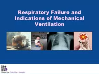
Respiratory Failure and the Need for Mechanical Ventilation. RES 300 Winter 2011. What is Respiratory Failure?. Defined as the inability to maintain oxygen delivery to the tissues or adequate removal of carbon dioxide from the body. Criteria PaO 2 < 60 mm Hg and/or PaCO 2 > 45 mm Hg

E N D
Respiratory Failure and the Need for Mechanical Ventilation RES 300 Winter 2011
What is Respiratory Failure? • Defined as the inability to maintain oxygen delivery to the tissues or adequate removal of carbon dioxide from the body. • Criteria • PaO2 < 60 mm Hg and/or • PaCO2 > 45 mm Hg • In individuals on room air at sea level • 36% hospital mortality
Hypoxemic Respiratory Failure (Type I) • Causes of hypoxemia • V/Q mismatch (most common cause) • Shunt • Alveolar hypoventilation • Diffusion impairment • Perfusion/diffusion impairment (rare) • Decreased inspired oxygen • Venous admixture
Clinical Presentation: V/Q Mismatch . • By definition patient has low PaO2 and SaO2 • Presents with • Nonspecific: dyspnea, tachycardia, tachypnea • Accessory muscle use (important sign) • Nasal flaring • Pedal edema (RF is cardiac in origin) • Cyanosis (peripheral or central) • Confusion to coma if CNS dysfunction
Clinical Presentation: V/Q Mismatch (cont.) • Auscultation • Bilateral wheezing • Bronchospasm, fluids, or upper airway disease • Breath sounds diminished bilaterally • Common finding with emphysema • Unilateral abnormalities important • Wheezing one lung may signify lesion • Absence of B/S one lung: collapse, effusion • Unilateral crackles: alveolar filling process
Shunt • Normal anatomic shunt ~2-3% of cardiac output • Pulmonary shunt occurs when there is NO ventilation to match perfusion • Always pathologic • Leads to hypoxemia as alveoli collapse or are filled with fluid or exudate • Atelectasis, pulmonary edema, pneumonia • Major difference between shunt and V/Q mismatch • V/Q mismatch responds to oxygen therapy unlike shunt which is refractory
Clinical Presentation: Shunt • Shunt presents very similar to V/Q match. • Shunt usually presents with a white radiograph. • ARDS would be the classic example. • Shunt is differentiated from V/Q mismatch by lack of response in PaO2 as the FIO2 is increased.
Diffusion Impairment • Most pronounced on exertion • Impairment can be caused by • Thickened/scarred: fibrosis, asbestosis • Alveolar destruction: emphysema • Pulmonary vascular abnormalities • Anemia, pulmonary emboli or hypertension • Clinical presentation depends on disease • Dry cough, fine bibasilar cracklespulmonary fibrosis • Jugular distention, edemapulmonary hypertension
Decreased Inspired Oxygen • Clinically uncommon • High altitude while mountain climbing • Airlines pressurized cabins but not to 1 atm. • Travelers with pulmonary disease may require supplemental oxygen or more supplemental oxygen than normal. • Signs and symptoms of hypoxemia may present. • Treat with oxygen
Venous Admixture • Decreased mixed venous oxygen • Clinically the patient’s lung must add more oxygen to the blood; the presence of pulmonary disease may prevent this. • Heart failure is most common cause. • Decreased cardiac output: tissues extract more oxygen • Clinically presents with signs and symptoms of CHF and/or underlying pulmonary disease
Differentiating Between Causes of Hypoxemic Respiratory Failure • Focus on three main causes: • Hypoventilation • Marked response to oxygen therapy • Normal P(A a)O2 • V/Q mismatch • Significant response to oxygen therapy • Increased P(A a)O2 • Shunt • Little or no improvement even on 100% O2 • Increased P(A a)O2
Deadspace • What is deadspace? • Ventilation without Perfusion • Total deadspace = anatomic + alveolar (all terminal respiratory units that are overventilated relative to their perfusion) • Anatomic = 1 cc/lb of ideal body weight • So, VE = Alveolar Ventilation + Deadspace Ventilation • VD/VT = PaCO2 – PECO2/PaCO2 • Normal is < 0.3 or 30% • Increased in Pulmonary Embolism • Increased in ARDS
Hypercapnic Respiratory Failure (Type II) • aka “pump failure” or “ventilatory failure” • An elevated PaCO2 results in uncompensated respiratory acidosis.
Ventilatory Failure: Decreased Ventilatory Drive • Ventilatory drive is most commonly diminished by: • Drug overdose or sedation • Brainstem lesions • Diseases of the CNS • Multiple sclerosis or Parkinson’s disease • Hypothyroidism • Morbid obesity • Sleep apnea • Clinical hallmark is bradypnea (<12 beats/min) and ultimately apnea.
Ventilatory Failure: Neurologic Diseases • CNS signals fail to reach the ventilatory muscles due to: • Spinal trauma • Motor neuron disease (ALS or polio) • Motor nerve disorders (GBS) • Neuromuscular junction disorders (MG or botulism) • Muscular diseases (MD, myositis)
Neurologic Diseases Clinical Presentation • Varied presentation • Drooling, dysarthria, weak cough – ALS • Lower extremity weakness progressing upward – GBS • Ocular muscle weakness, ptosis, diplopia, dysphagia – Myasthenia gravis • Different clinical presentations, yet they commonly result in respiratory muscle fatigue and ventilatory failure (elevated CO2).
Ventilatory Failure: Increased Work of Breathing • Ventilatory failure may occur if the imposed workload cannot be overcome. • Most commonly occurs secondarily to • Increased VD/VT in COPD • Elevated Raw in asthma • Both cause intrinsic PEEP, which generates excessive work of breathing (WOB) • May also be caused by • Pneumothorax, rib fractures, pleural effusions • Hypermetabolic states such as burns
Increased Work of Breathing Clinical Presentation • Rapid shallow breathing is an indication of impending ventilatory failure. • Shallow breathing increases the VD/VT ratio and results in hypercapnia. • Diminished B/S in a young asthmatic is ominousnot moving adequate air • Irritability, confusion, and coma are signs of worsening hypercapnia. • Muscle tremors and papilledema
Chronic Respiratory Failure (Type I and Type II) • Over months and years, acute respiratory failure will become a chronic condition. • Body develops compensatory mechanisms. • Chronic type I failure (hypoxemic) • Polycythemia and oxy-Hb shift to right • Cerebral blood flow enhanced by increased PaCO2 • Chronic ventilatory failure (hypercapnic) • Renal response: retain HCO3 to normalize pH • Will be incomplete but will raise pH toward normal
Acute-on-Chronic Respiratory Failure • Chronic failure complicated by acute failure. • This is most commonly brought about by • Bacterial or viral infections • CHF • Pulmonary embolus • Chest wall dysfunction • Medical noncompliance • Key: Treat aggressively to prevent further exacerbations.
Complications of Acute Respiratory Failure • Complications add significantly to morbidity and mortality. • In ARDS, more patients die of complications (sepsis, MSOF) than of the original disease. • Emboli, barotrauma, and infection are common. • Nonpulmonary complications include • Cardiac: arrhythmias, hypotension • Gastrointestinal: hemorrhage, dysmotility • Renal failure and/or positive fluid balance
Clinical Presentation of Acute Respiratory Failure • Respiratory muscle fatigue presents • Tachypnea: cardinal sign of increased WOB • Worsening fatigue RR starts falling, bradypnea occurs and, with progression, apnea • Respiratory alternans • With full ventilatory failure • ABG: hypercapnia with acidosis
Indications for Ventilatory Support Goal of Mechanical Ventilation • Supportive therapy until underlying problem resolves OR • Provide long-term support for patients with chronic ventilatory failure • Support will be aimed at the patient’s specific needs • A pneumonia patient’s ventilatoryneeds will differ markedly from that of a patient with a C1 spinal cord injury.
Indications for Ventilatory Support (cont.) • Values that indicate mechanical ventilation • handout
Assessment of Respiratory Muscle Weakness • Commonly occurs in neuromuscular disease (NMD) patients, but also COPD and kyphoscoliosis. • Tests to assess respiratory muscle strength • MIP of >20 is inadequate, but watch trends. • In NMD the trend of MIP becoming less negative indicates impending ventilatory failure
Respiratory Muscle Weakness, Fatigue, and Failure • Weakness, fatigue, and failure overlap and may result in acute or chronic failure. • Excessive WOB is the most common cause of respiratory muscle fatigue and failure to wean from MV. • Imposed WOB in ventilated patients is due to: • ETT • Ventilator circuit • Auto-PEEP • Disease process
Are there alternatives? • Noninvasive positive-pressure ventilation provides support without intubation. • Methods • CPAP- one continuous pressure • used for oxygenation and stinting airway in OSA • BiPap- one pressure for inspiration and one for expiration • used for ventilation, oxygenation and stinting airway in OSA
NPPV • Indications • Allows patient to be supported while other therapeutic maneuvers can help the patient while decreasing the WOB • Disorders that can be rapidly reversed • Clinical situations that may respond to NPPV • Acute exacerbations of COPD (good evidence) • Cardiogenic pulmonary edema (reversibility) • Acute asthma (use is controversial) • Acute type I failure: improved PaO2/FiO2 ratio but no patient improvement outcomes (i.e., LoS, M/M) • Chronic type II failure particularly NMD: improves and prolongs life and cognitive function and reduces pneumonia and hospitalization
What if our problem is oxygenation alone? • Can try CPAP and/or Vapotherm